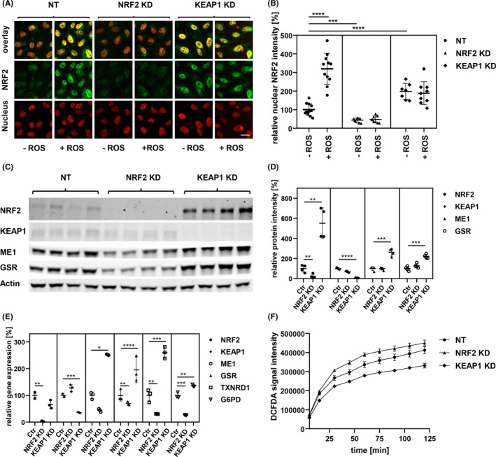
FIGURE 1.

Melanoma cell lines were modified by siRNA to activate or inactivate the NRF2/KEAP1 regulation system. (A) Immunofluorescence images of WM793b cells with siRNA treatments, nucleus staining by DAPI, ROS stimulation with 5 µg/ml of Pyocyanin for 6 h, Scale bar: 10 µm. (B) Quantification of color intensity of NRF2 secondary antibody fluorescence in nuclear area, indicated by DAPI staining (unpaired t‐test; ***p‐value < 0.001; ****p‐value < 0.0001). (C) Western blot of WM793b protein extract 48 h after siRNA transfection, NRF2 and KEAP1 as siRNA targets, ME1 and GSR as targets of NRF2 transcription factor, β‐Actin as loading control. (D) Quantification of Western blot shown in C (unpaired t‐test; **p‐value < 0.01; ***p‐value < 0.001; ****p‐value < 0.0001). (E) qPCR of WM793b cDNA 24 h after siRNA transfection, NRF2 and KEAP1 as siRNA targets, ME1, GSR, TXNRD1 and G6PD as targets of NRF2 transcription factor (unpaired t‐test; *p‐value < 0.05; **p‐value < 0.01; ***p‐value < 0.001; ****p‐value < 0.0001). (F) ROS quantification by DCFDA fluorescence assay over 2 h with 10 mM Hydrogen peroxide (H2O2), in WM793b 48 h after siRNA transfection
