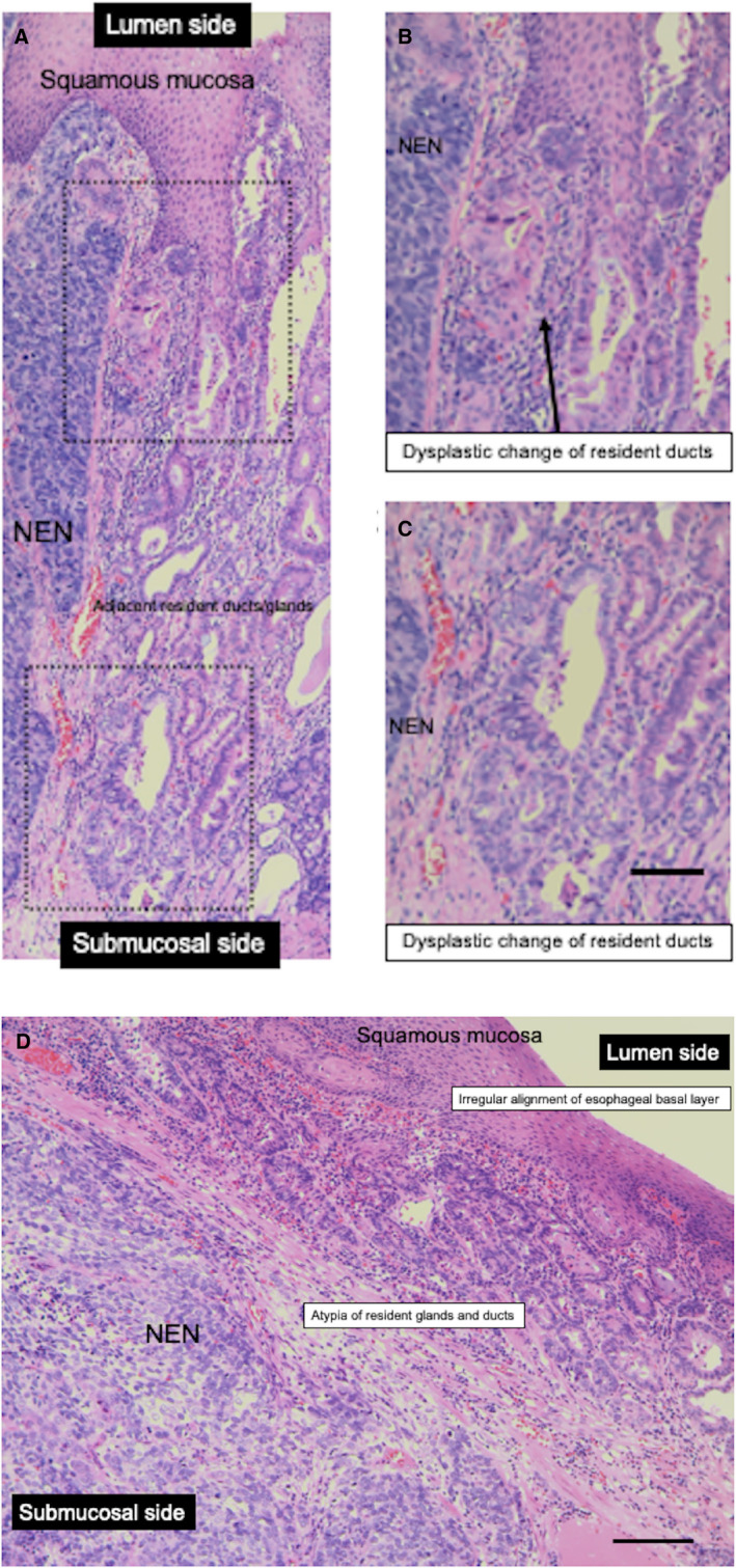
FIGURE 4.

Histological transformation of resident ducts/glands in a representative esophageal NEN sample. (A) NEN‐adjacent dysplasia of ducts in a resected esophageal NEN sample (in the esophagogastric junction, the NEN tumor invades submucosa) (case 10 in Table 1). (B), (C) High‐power view of NEN‐adjacent transformation of resident ducts in a resected esophageal NEN sample. (D) NEN‐adjacent atypia of resident glands and ducts in a resected esophageal NEN sample. Black bar indicates 100 μm. NEN, neuroendocrine neoplasm
