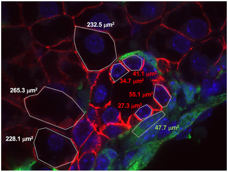
Fig 6. Detail image of the lumen with indicated cell surface areas.
White numbering: hepatoblasts, red numbering: cholangiocytes, green numbering: mesenchyme. The picture is from an E18.5 mouse liver expressing eYFP in the mesenchyme. Red membrane staining of hepatoblasts and cholangiocytes: E-cadherin; green staining of mesenchyme: eYFP.

