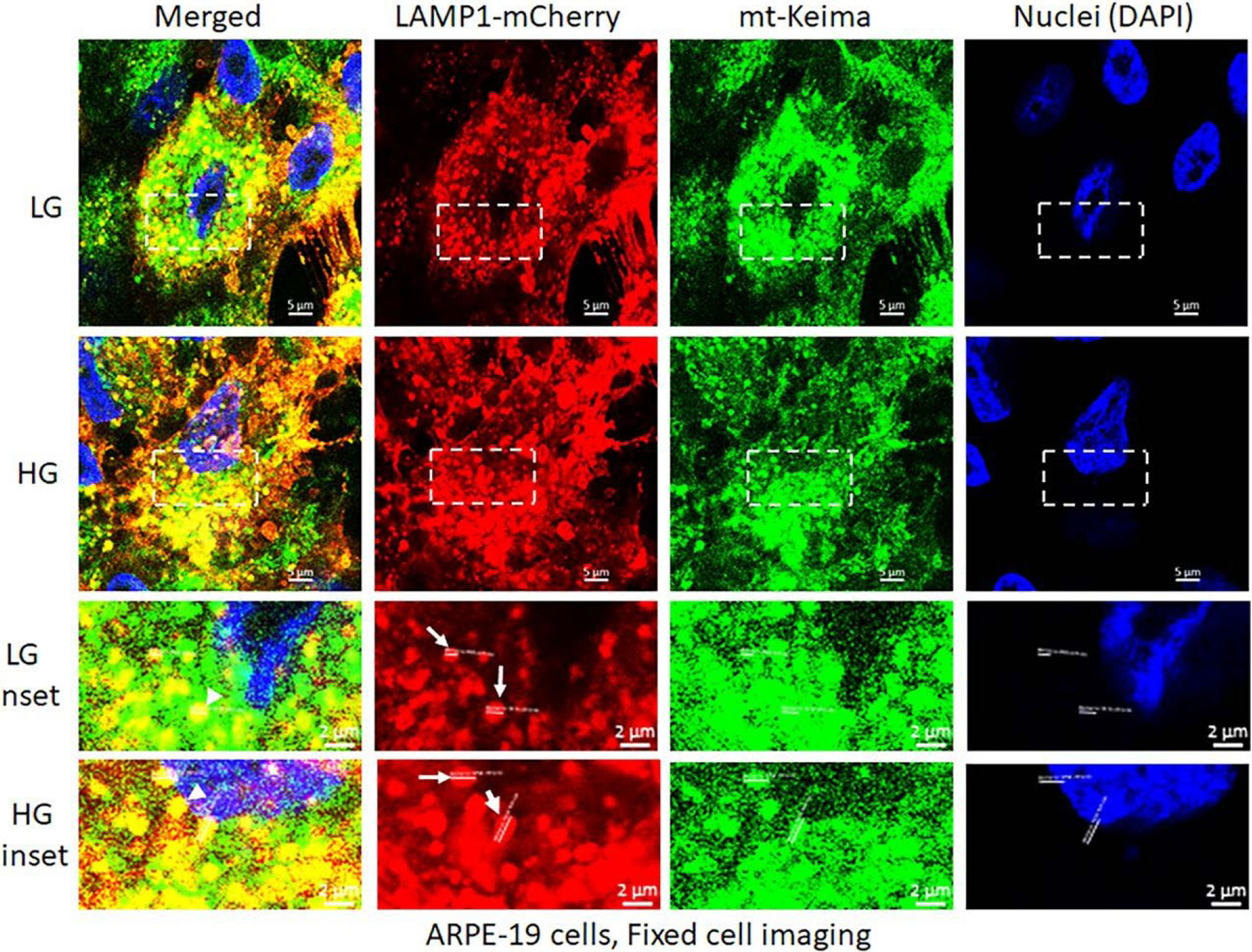
Figure 2:

High glucose induces mitophagic flux and lysosome enlargement in ARPE-19 cells. Transduction of mt-Keima and LAMP1-mCherry bearing adenovirus vectors in APRE-19 cells were described previously [33]. Mt-Keima and LAMP1-mCherry vectors were transfected together in ARPE-19 cells and incubated with LG (5.5mM) or HG (25mM glucose) for 5 days, then the cells were fixed, mounted on DAPI containing mounting medium to stain nuclei and imaged with a Zeiss Confocal microscopy at 630x magnification. The images were analyzed by Zen 3.0 blue software and compile in Adobe Photoshop. Under these experimental conditions, mt-Keima emits green both in mitochondria and lysosomes while LAMP1-mCherry emits red in lysosomes. With HG treatment, lysosome sizes are ~1.5- to 2-folds larger than in LG (mCherry, white arrows in insets) while there are also more yellow mito-lysosomes in HG versus LG (green and red combination, arrowheads in insets). A representative of three experiments is shown here.
