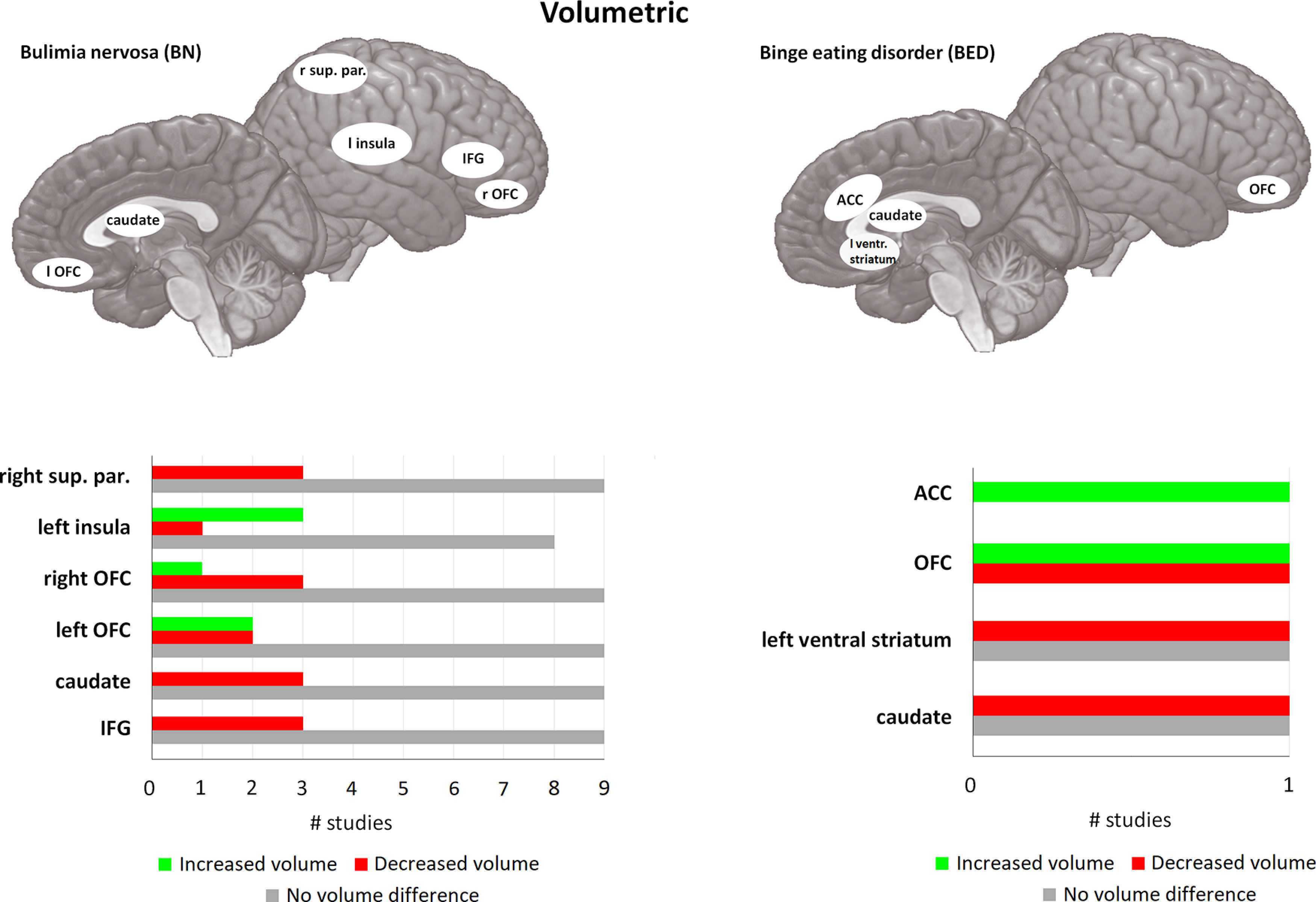
Figure 4.

Volumetric differences in BN and BED compared with matched controls. For each area, the bar graph indicates the number of studies that found a reduction in volume (= red), or an increase in volume (= green), and the studies that found no difference in activity (= gray). For BN, 10 of the 14 included studies found at least one brain area that was significantly different compared with HC. Four studies did not find any significant differences between BN and HC. caudate = caudate nucleus, OFC = orbitofrontal cortex, sup. par. = superior parietal cortex, IFG = inferior frontal gyrus, ventr. striatum = ventral striatum, ACC = anterior cingulate cortex. If no indication of lateralization is given (either left or right), differences are observed bilateral. For the left part (BN) of this figure only, areas with one study indicating differences are not displayed, because of the large number of areas found in BN. For the right part (BED) all studies are displayed. For a full overview for differences in BN, please see Table 2.
