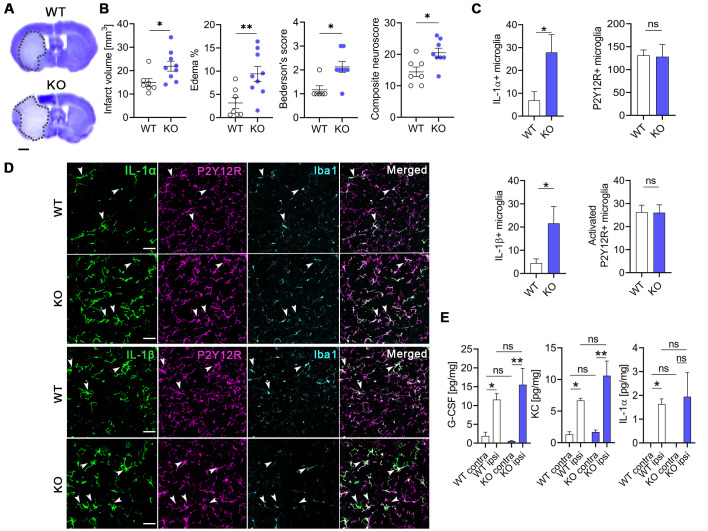
Fig 6. Deletion of microglial NKCC1 increases infarct volume, brain edema, and worsens neurological outcome after MCAo.
(A, B) Microglial NKCC1-deficient mice (KO) show larger infarct volume as assessed on cresyl violet-stained brain sections and more severe neurological outcome compared to WT mice. (C, D) Microglial NKCC1 deletion results in higher levels of IL-1α and IL-1β 24 hours after MCAo. (E) Cytokine levels in the cortex do not differ 8 hours after MCAo in KO mice compared to WT. (A) Scale: 1 mm. (B) Unpaired t test, N (WT) = 7, N (KO) = 9; *: p < 0.05, **: p < 0.01. (C) Mann–Whitney test, *: p < 0.05; N (WT) = 7, N (KO) = 8. (D) Scale: 50 μm. (E) Kruskall–Wallis test followed by Dunn’s multiple comparison test; N = 6/group. *: p < 0.05, **: p < 0.01. Data underlying this figure can be found in S1 Data. KO, knockout; MCAo, middle cerebral artery occlusion; ns, not significant; WT, wild type.

