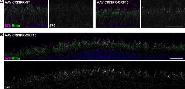
Fig. 4. Correction of RPGR-ORF15 in AAV-treated rd9 mice.
A Confocal images of representative AAV-treated rd9 retinal cross sections demonstrating rescue. Expression of RPGR-ORF15 is observed only in ORF15-targeted (AAV CRISPR-ORF15) but not non-targeted (AAV CRISPR-NT) retina, as indicated by a subset of cells positive for RPGR-570 signal at the connecting cilium. The ciliary rootlet is stained with Rootletin (Rtltn). Right panels show desaturated 570 channel to better visualize positive signal. B Lower magnification image of staining depicted in panel (A) to better show broad distribution of correction in treated retina. Lower panel shows desaturated 570 channel. Scale bars indicate 20 μm. Injections were performed in 3–6 week old rd9 mice, and tissues were collected and analyzed 8–12 weeks post injection.

