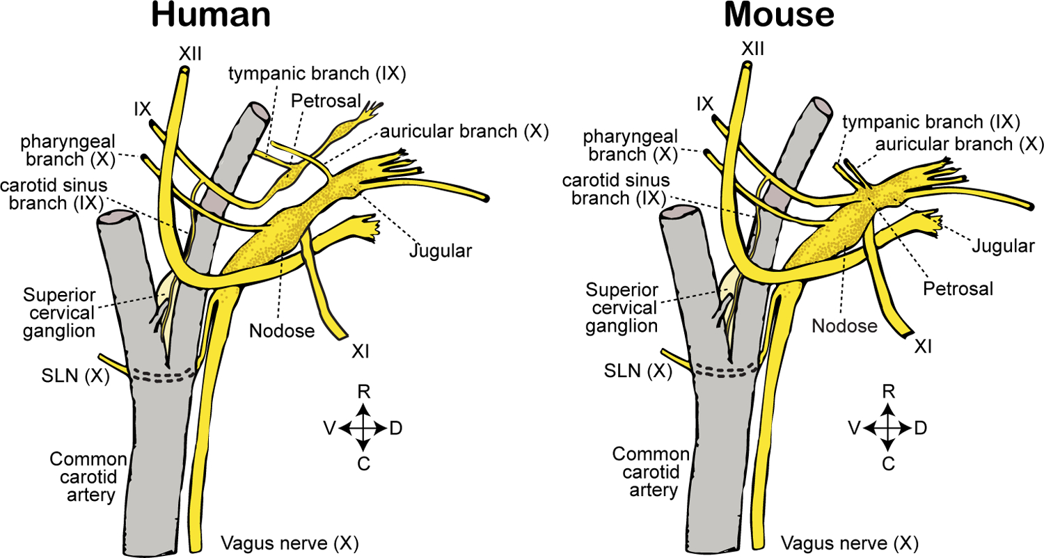
Figure 2. Comparative anatomy of cranial ganglia and branches in mice and human.

Sensory ganglia and branches of cranial nerves IX (glossopharyngeal) and X (vagus) are depicted in relation to nearby anatomical structures. Adult mice display a fused nodose-petrosal-jugular superganglion (left); in some animals, the superior cervical ganglion (here depicted separately) is additionally fused into the superganglion. In humans (right), vagal and petrosal ganglia are distinct anatomical structures. SLN: superior laryngeal nerve. R: rostral. C: caudal. D: dorsal. V: ventral.
