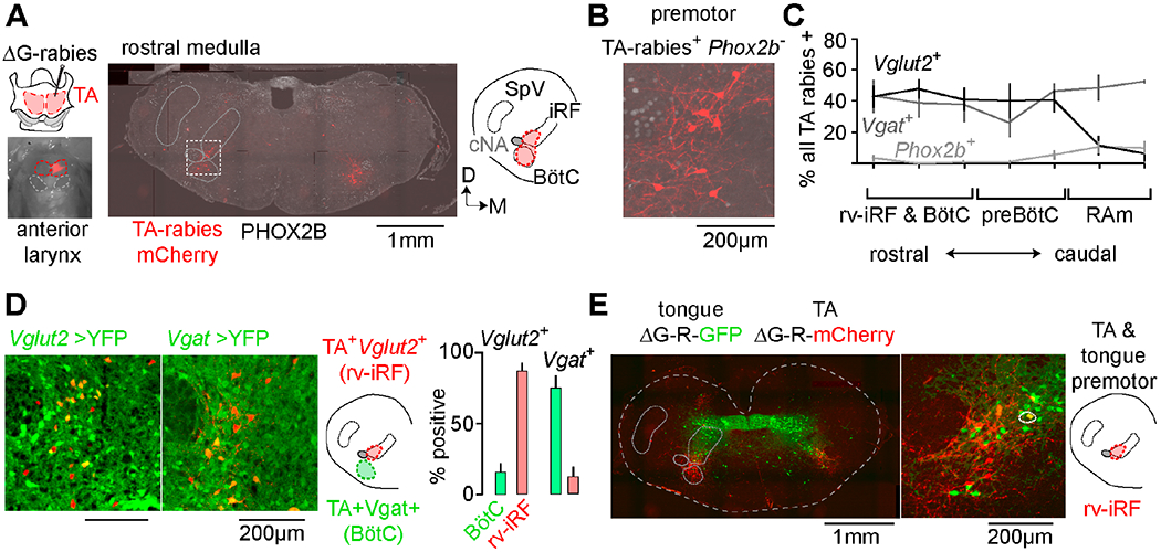
Figure 3. The medullary brainstem contains a cluster of premotor neurons for sound production and articulation.

A, Modified rabies (ΔG-Rabies-mCherry) and helper (HSV-G) viruses co-injected into TA muscle. Middle, coronal section of rostral medulla showing distribution of rabies-mCherry+ TA premotor neurons (red) and Phox2b (gray). Right, schematic illustrating the position of the intermediate reticular formation (iRF) and BötC, cNA, and spinal trigeminal nucleus (SpV). Dorsal; Medial. B, Boxed area in A showing a TA premotor neuron cluster (Phox2b-neg.) directly medial to cNA. C, Quantification of mean % ± SEM of rabies+ neurons that were Vglut2+ (black, n=3), Vgat+ (gray, n=3), or Phox2B+ (TA motor, pale gray, n=6) across the breathing control centers (rv-iRF/BötC, preBötC, and RAm). D, rv-iRF and BötC TA premotor neurons in Vglut2-Cre→YFP (n=3) and Vgat-Cre→YFP mice (n=3). Those medial to cNA are Vglut2+ (rv-iRF) and in BötC are Vgat+. Right, quantification. E, Coronal section showing the distribution of ΔG-rabies-mCherry TA premotor neurons and ΔG-rabies-GFP tongue premotor neurons. Middle, magnification of rv-iRF showing spatial overlap of premotor neurons. Circled neuron is double positive. See also Figures S3 and S4.
