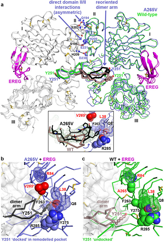
FIGURE 4. A265V mutation optimises dimer arm docking in EREG-induced dimers.
a. Overlay of EREG-induced sEGFRWT and sEGFRA265V dimers. The left-hand molecule is grey, and the right-hand molecule slate blue (A265V) or green (WT). The dimer arm of the left-hand molecule is black (A265V) or salmon (WT). The insert shows an unbiased ∣Fo∣-∣Fc∣ polder OMIT map18 at 3.5 Å resolution (contoured at 2σ), calculated using A265V data and omitting L243-P257 from the model.
b. Close-up of (black) dimer arm docking in the unique site (slate blue) formed in the A265V-mutated EREG-induced dimer. The mutated V265 is red.
c. Close-up of undocked dimer arm (salmon) in EREG-induced WT sEGFR dimers.

