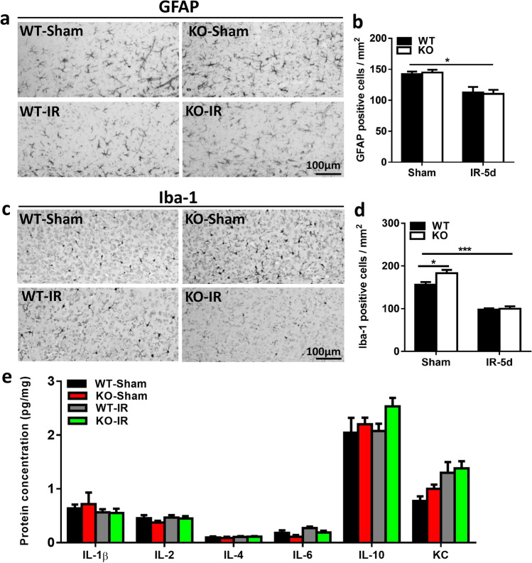
Fig. 3.
Astrocyte and microglia changes in the subcortical white matter after irradiation a Representative images of GFAP-labeled cells in the subcortical white matter. b Quantification of GFAP-labeled cells in the subcortical white matter. c Representative Iba-1 immunostaining in the subcortical white matter. d Quantification of Iba-1–labeled cells in the subcortical white matter. e The protein levels of IL-1β, IL-2, IL-4, IL-6, IL-10, and KC in the cortical tissue at 5 days after irradiation as detected by Luminex assay in the Atg7 KO and WT pups. n = 7/group for the immunostaining and Luminex assay. *p < 0.05, ***p < 0.001.

