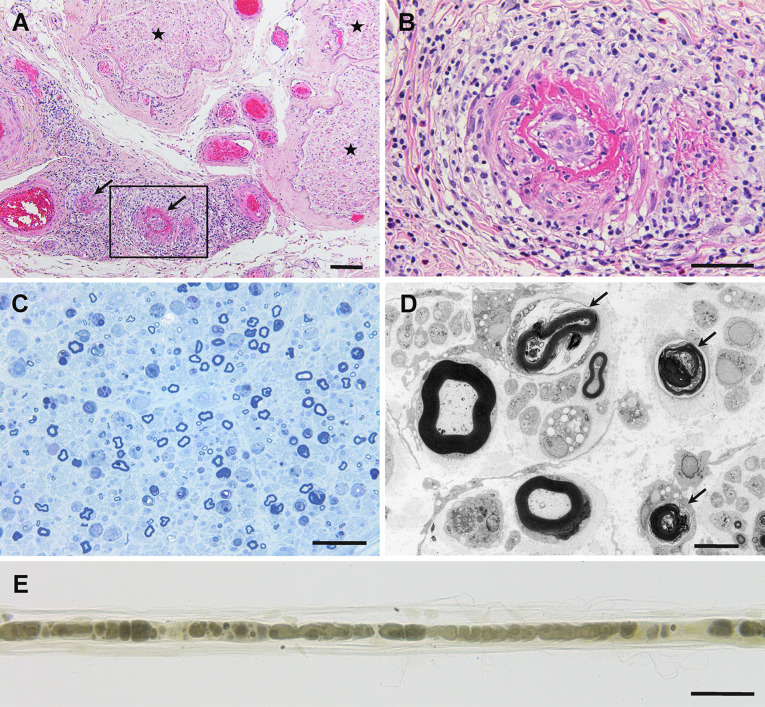
Fig. 1.
Representative photographs showing neuropathy in anti-neutrophil cytoplasmic antibody-associated vasculitis. Cross-sections (a–d) and a teased-fiber preparation (e) of sural nerve biopsy specimens obtained from patients with microscopic polyangiitis. a Epineurial vessels indicated by arrows show fibrinoid necrosis. Massive inflammatory cell infiltration was observed around these vessels. The endoneurium, where nerve fibers are located, is indicated by asterisks. b A high-powered view of the region in the box in (a). c The density of myelinated fibers is reduced. d Degeneration of myelinated fibers is evident (arrows) via electron microscopy. e Teased-fiber preparations also demonstrated myelinated fiber degeneration. Hematoxylin and eosin staining (a, b), toluidine blue staining (c), uranyl acetate and lead citrate staining (d), and osmium staining (e). Scale bars = 100 μm (a), 50 μm (b, c, e), and 5 μm (d)

