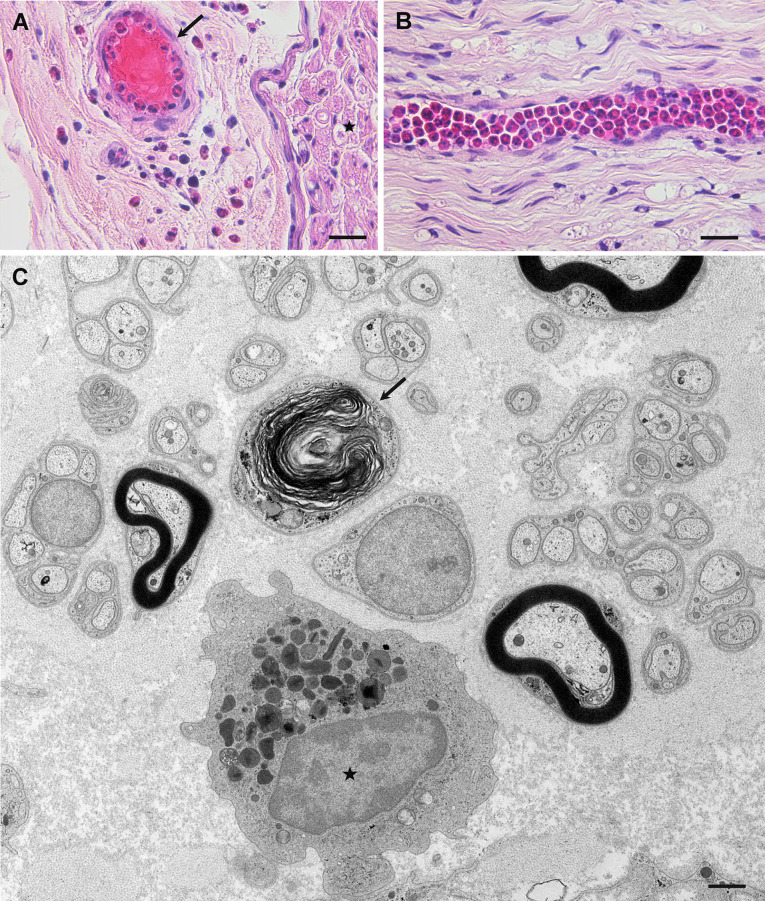
Fig. 3.
Pathological findings of eosinophilic granulomatosis with polyangiitis. Sural nerve biopsy specimens obtained from patients negative for anti-neutrophil cytoplasmic antibody. Cross-sections (a, c) and a longitudinal section (b). a Infiltration of eosinophils into the extravascular space in the epineurium is observed. Eosinophils are seen in the lumen of a vessel (arrow). An endoneurium, where the nerve fibers are located, is indicated by an asterisk. b Many eosinophils are packed inside an endoneurial vessel. c An eosinophil indicated by an asterisk is located in the extravascular space of the endoneurium. The arrow indicates a degenerated myelinated fiber. Hematoxylin and eosin staining (a, b); uranyl acetate and lead citrate staining (c). Scale bars = 20 μm (a, b) and 1 μm (c)

