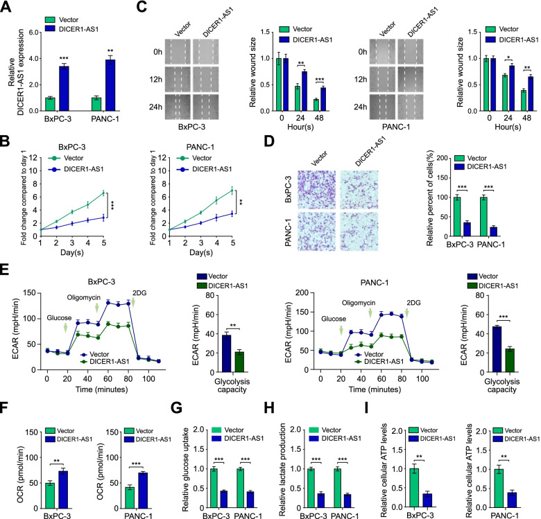
Fig. 2.
DICER1-AS1 inhibits glycolysis, proliferation and metastasis of PC cells. A Real-time PCR analysis of DICER1-AS1 level in BxPC-3 and PANC-1 cells after transfected with the pcDNA-DICER1-AS1 plasmid (DICER1-AS1) and negative control (Vector), respectively. B The proliferative capability was performed by MTT assay in BxPC-3 and PANC-1 cells transfected with pcDNA-DICER1-AS1 (DICER1-AS1) or empty vector control (Vector). C Migration ability was assessed by wound healing assay in BxPC-3 and PANC-1 cells transfected with pcDNA-DICER1-AS1 (DICER1-AS1) or empty vector control (Vector). D Transwell assay was applied to assess the invasion ability of those cells transfected indicated above. E The change of ECAR levels in BxPC-3 and PANC-1 cells transfected with vector or DICER1-AS1 (n = 3). The glucose (10 mmol·L−1), oligomycin (2 μmol·L−1), or 2-deoxyglucose (2-DG, 50 mmol·L−1) were used at indicated points. F Seahorse extracellular flux assay showing the oxygen consumption rate (OCR) in BxPC-3 and PANC-1 cells transfected with vector or DICER1-AS1 (n = 3). G The glucose uptake levels in BxPC-3 and PANC-1 cells transfected with control or DICER1-AS1 vectors (n = 3). H The relative lactic acid levels were detected in BxPC-3 and PANC-1 cells transfected with control or DICER1-AS1 vectors (n = 3). I The ATP levels in BxPC-3 and PANC-1 cells transfected with control or DICER1-AS1 vectors (n = 3). All data were presented as means ± SD of at least three independent experiments. Values are significant at aP < 0.05, bP < 0.01 and cP < 0.001 as indicated

