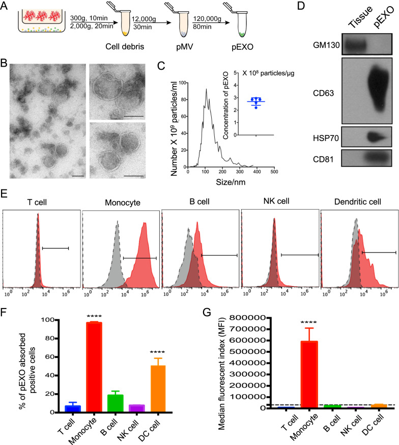
Fig. 1.
Characterization of pEXOs and interaction by different immune cell populations. A Schematic illustration of the sequential centrifugation to isolate exosomes from conditioned medium of human placenta explant. B Transmission electron microscopy images of pEXO, Scale bar = 100 nm. C NTA of the isolated pEXO. Particle concentration of pEXO is approximately 2.75 × 10⁸ particles/μg exosome protein. The mean size is 113 nm. D Western blot analysis demonstrates the presence of exosome markers CD63, HSP70, CD81 and the absence of GM130 (Golgi marker). E Flow cytometric analysis demonstrating the interaction of fluorescently labelled pEXO with different immune cell populations after 24 h. pEXO were mainly interacted with CD14+ monocytes. B cells and dendritic cells have a mild interaction with pEXO, while T cells and NK cells have barely no interaction. F Percentage of Carboxy-fluorescein succinimidyl ester (CFSE)-labelled pEXO positive cells and G Median fluorescent index (MFI) in different immune cell populations. Data are expressed as mean ± SD (n = 4), *p < 0.05, **p < 0.01, ***p < 0.001, ****p < 0.0001 compared to the control group

