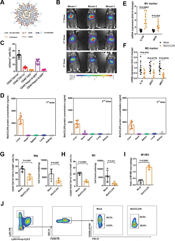
Fig. 11.
Dual blockade of CCL2 and CCL5 via LNP-mediated mRNA delivery of BisCCL2/5i polarizes macrophage M1 phenotype and reduces the immunosuppression in the TME. A Schematic of the mRNA-loaded LNPs. B In vivo transfection of Luc mRNA-LNPs after repeated administration (i.v., every 4 days, in total 3 doses). The luciferase was injected into mice 6 h post administration of Luc mRNA-LNPs, followed by measuring luc bioluminescence signal using IVIS imaging, n = 3. C The quantification of mCherry-positive cells expressed in murine orthotopic HCC tumor tissue 6 h after injection of mCherry mRNA-LNPs (mCherry mRNA: 0.5 mg kg − 1). mRNA is mainly expressed in monocytes (CD45 + CD11b+) and tumor cells (Hepa1–6-GFP+) (n = 8). D BisCCL2/5i expression in different organs 6 h after each administration of BisCCL2/5i mRNA-LNPs (mRNA: 1 mg kg − 1, i.v., 3 days apart), n = 6. The BisCCL2/5i mRNA was mainly expressed in liver tissue and repeated administration resulted in comparable protein level. E, F mRNA expression of classic M1 (E) and M2 (F) markers in HCC tumor tissues 48 h after systemic administration of formulated LNPs as a dose corresponding to 1 mg kg − 1 mRNA (Mock, HcRed mRNA). Each data point is an individual sample (n = 9); one-way ANOVA and Tukey’s multiple comparisons test. Change of the immunocellular composition in HCC TME 48 h following Mock mRNA-LNPs and BisCCL2/5i mRNA-LNPs treatments (mRNA: 1 mg kg − 1), measured by flow cytometry (n = 4; unpaired two-tailed Student’s t-test). G, H The percentage and cell counts of macrophages (G) and their M2 subtype (H) in total immune cells. I, J Representative flow dots of M1- and M2-phenotype macrophages (I) and ratio of M1/M2 (J). MΦ, macrophages (CD45 + CD11b + CD11c − Ly6C − Ly6G − F4/80+); M2, M2-phenotype macrophages (CD206+). Data are represented as the mean ± s.d. [164]

