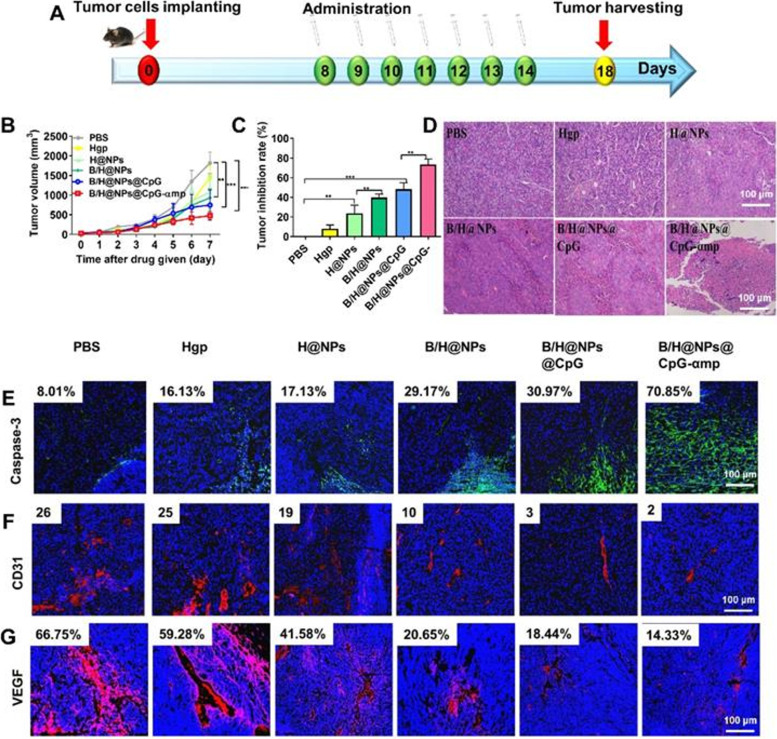
Fig. 12.
Effective anti-tumor and tumor microenvironment remodeling after treatment with different nano-complexes in the B16 tumor model. A Schematic illustration of the time sequence of administration of nano-complex to tumor-bearing mice. B Tumor volume from mice that received iv infusion containing different nano-complexes. C Tumor inhibition fractions after receiving iv infusion of various nano-complexes formulations. D Evidence of necrosis in tumors after treatment with different nano-complexes by hematoxylin and eosin (H&E) staining. E Caspase-3 analysis of tumor tissue indicating apoptotic cells by immunofluorescence in frozen tumor sections. F The number of vessels per image field is identified by CD31 label after treatment with different nano-complexes. G VEGF labeled by immunofluorescence indicates the quality of pro-angiogenesis secretion per image field after treatment with different nano-complexes. The data were analyzed by automatic multispectral imaging system (PerkinElmer Vectra II). Scale bar: 100 μm. Three mice were analyzed in every group (n = 3), and one representative image per group is displayed. Data are the mean ± SEM and representative of three independent experiments. Differences between two groups were tested using an unpaired, two-tailed Student’s t-test. Differences among multiple groups were tested with one-way ANOVA followed by Tukey’s multiple comparison. Significant differences between groups are expressed as follows: *P < 0.05, **P < 0.01, or ***P < 0.001 [169]

