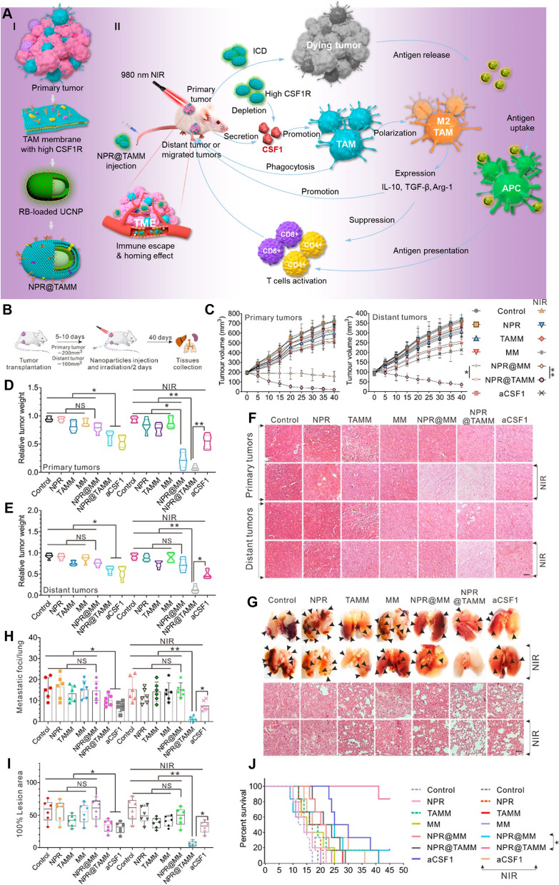
Fig. 15.
A Schematic illustration of the tumor-associated-macrophage-membrane-coated up conversion nanoparticles for improved photodynamic immunotherapy. B-J In vivo antitumor therapeutic effects. B Schematic illustration of 4 T1 tumor model establishment and the therapeutic regimen. C Tumor growth curves for primary tumor and distant tumor. D Tumor weight for primary tumor. E Tumor weight for distant tumor. F Histological analysis of H&E staining for primary and distant tumor. G Photographs show representative external views of lung with the histological analysis of H&E staining. Arrows indicate focal tumor nodules on lung surfaces. Scale bar = 100 μm. (H, I) Graphs show the quantification of metastatic foci (H) and lesion area (I) in the different treatment groups from part f. J The survival curve of tumor-bearing mice calculated by Kaplan−Meier estimate. Data are means ± SD. *P < 0.05; **P < 0.01. NS, no significance. n = 6/group [203]

