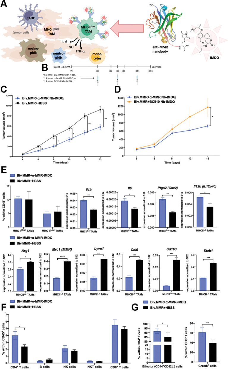
Fig. 6.
A A well-defined protein-drug conjugate of anti-MMR nanobody with TLR 7/8 agonist IMDQ. The anti-MMR Nb-IMDQ conjugate allows triggering of TLR7/8 specifically of MMR high macrophages, with aim to repolarize these cells into a pro-inflammatory anti-tumoral state, resulting in reduced tumor growth. B-G Anti (α)-MMR Nb-IMDQ therapy delays tumor progression and reprograms TAMs to more M1 phenotype. B LLC-OVA-bearing C57BL/6 mice were injected on day 5, 8, and 11 after cancer cell inoculation with appropriate treatment and mice were sacrificed on day 13. C LLC-OVA bearing mice received α-MMR Nb-IMDQ or HBSS, co-injected with fivefold molar excess of bivalent α-MMR Nb (Biv.MMR) and tumor volumes were measured on day 4, 6, 8, 10, 12, and 13 after cancer cell inoculation. D LLC-OVA bearing mice received α-MMR Nb-IMDQ or BCII10 Nb-IMDQ, co-injected with fivefold molar excess of Biv.MMR, tumor volumes were measured on day 6, 8, 11, 12, and 13 after cancer cell inoculation. p-values are calculated using a two-way ANOVA and significant differences are marked by *: p ≤ 0.05. E The percentage of MHC-II high and MHC-II low TAMs within hematopoietic (CD45+) cells of LLC-OVA tumors is shown as mean ± SEM of n = 4. MHC-II low TAMs were sorted from pools of tumor cell suspensions of each individual experimental group and qRT-PCR analysis was performed for technical triplicates to quantify expression of several M1 and M2-associated genes normalized to ribosomal protein S12 expression. F Percentage of CD4+ T cells, B cells, NK cells, NKT cells, and CD8+ T cells within the hematopoietic (CD45+) cells is shown as mean ± SEM of n = 4, p ≤ 0.05. G Percentage of effector (CD44 + CD62L−) cells within CD4+ T cells and Gzmb+ cells within CD8+ T cells is shown as mean ± SEM. p ≤ 0.05; **: p ≤ 0.01 [147]

