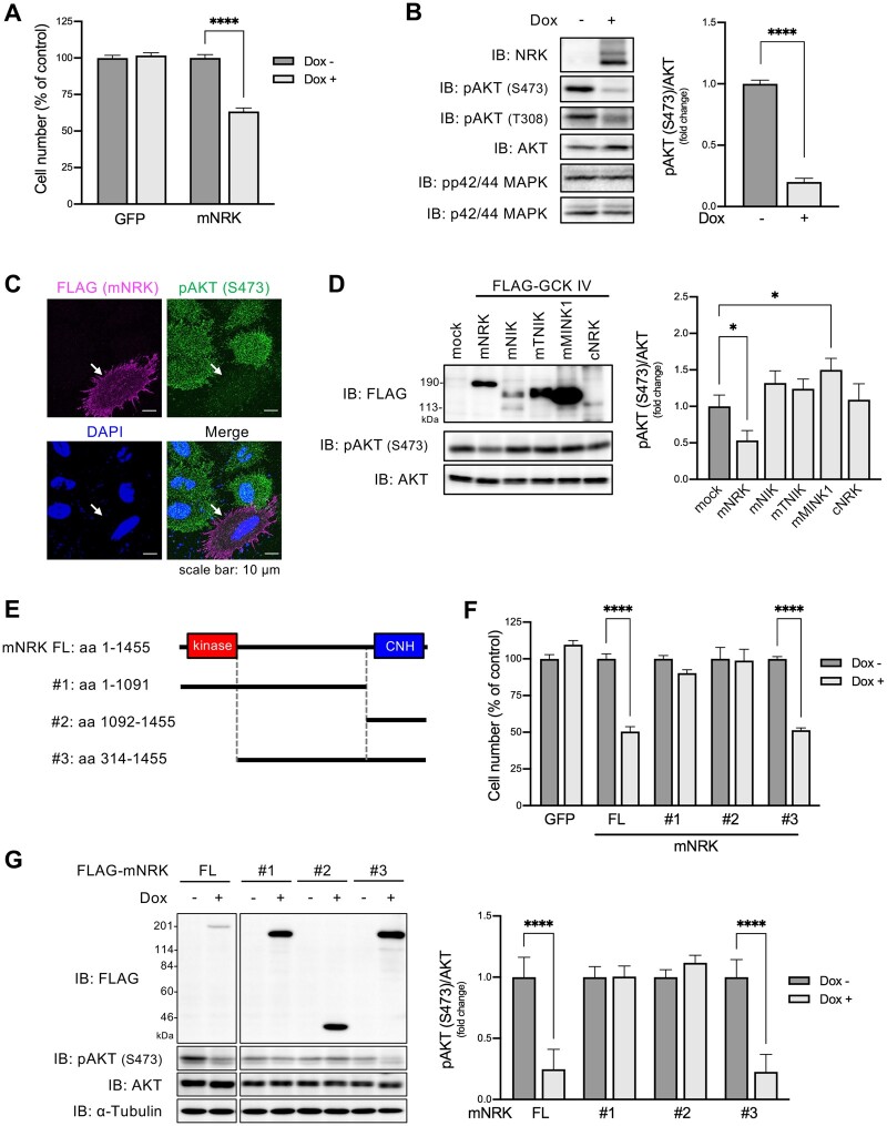
Fig. 3.
Effects of mNRK on cell proliferation and AKT signaling and the corresponding responsible regions of mNRK. (A) Effects of mNRK on cell proliferation. We generated Flp-In T-REx 293 cell lines that expressed mNRK and GFP in doxycycline (dox)-dependent manner. The cells were treated with dox (1 μg/ml) for 2 days and then subjected to MTT assay. The graph shows the mean ± SD of three independent experiments. ****P ≤ 0.0001 (unpaired two-tailed Student’s t-test). (B) Effects of mNRK on AKT signaling analyzed by immnoblotting. Cells prepared by a method similar to that shown in (A) were subjected to immunoblotting. Densitometric analyses were performed, and phosphorylation levels of AKT (S473) were normalized to protein levels of AKT. The graph shows the mean ± SD of three independent experiments. ****P ≤ 0.0001 (unpaired two-tailed Student’s t-test). (C) Effects of mNRK on AKT signaling analyzed by immnostaining. HeLa cells expressing FLAG-tagged NRK were stimulated with EGF (100 ng/ml) for 10 min and subjected to cell staining with anti-FLAG antibody (magenta), anti-phospho AKT (S473) antibody (green), and DAPI (blue). Arrows indicate NRK-positive cells. Scale bar, 10 µm. (D) Effects of the other GCK IV family members (mNIK, mTNIK, and mMINK1) and cNRK on AKT signaling. HEK293 cells were transfected with plasmids encoding mNRK, mNIK, mTNIK, mMINK1, and cNRK. The lysates were subjected to immunoblotting, followed by densitometric analyses. The graph shows the mean ± SD of three independent experiments. *P ≤ 0.05 (one-way analysis of variance [ANOVA], followed by Dunnett’s multiple comparisons test). (E) Schematic structures of the truncated forms of mNRK used in (F) and (G). (F) Effects of the truncated forms of mNRK on cell proliferation. We generated Flp-In T-REx 293 cell lines that expressed the truncates in a dox-dependent manner. The cells were treated with dox (1 μg/ml) for 2 days and then subjected to the MTT assay. (G) Effects of the truncated forms of mNRK on AKT signaling. Cells treated by a method similar to that shown in (F) were subjected to immunoblotting, followed by densitometric analyses. The graphs show the mean ± SD of three independent experiments. ****P ≤ 0.0001 (unpaired two-tailed Student’s t-test).

