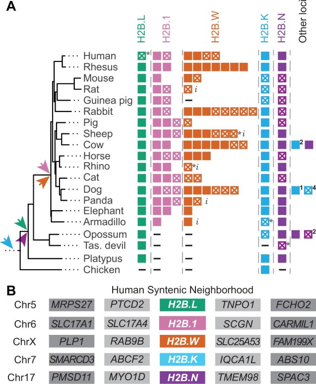
Fig. 3.

Retention and synteny of identified H2B variants in mammals. (A) A schematic representation of H2B variants and paralogs, along with a species tree of selected representative mammals and a nonmammalian outgroup, chicken, (Bininda-Emonds et al. 2007). Colors distinguish H2B variants as in figure 1A. Filled boxes represent intact ORFs and empty boxes with a cross represent interrupted ORFs (inferred pseudogenes). An asterisk (*) indicates pseudogenization by a single-nucleotide change which could either be sequencing error or a true mutation. A dash (–) indicates absence of histone within the syntenic location, and an “i” indicates incomplete sequence information. Colored arrows indicate the predicted origin of each H2B variant. Copies of variants found outside the syntenic neighborhood are shown as “other loci” with number of intact ORFs and pseudogenes indicated. (B) A schematic of the shared syntenic genomic neighborhoods of each H2B variant in the human genome. All H2B variants in (A) were present in the same syntenic location across mammals (see supplementary fig. S8, Supplementary Material online, for detailed syntenic analyses) except “other loci.”
