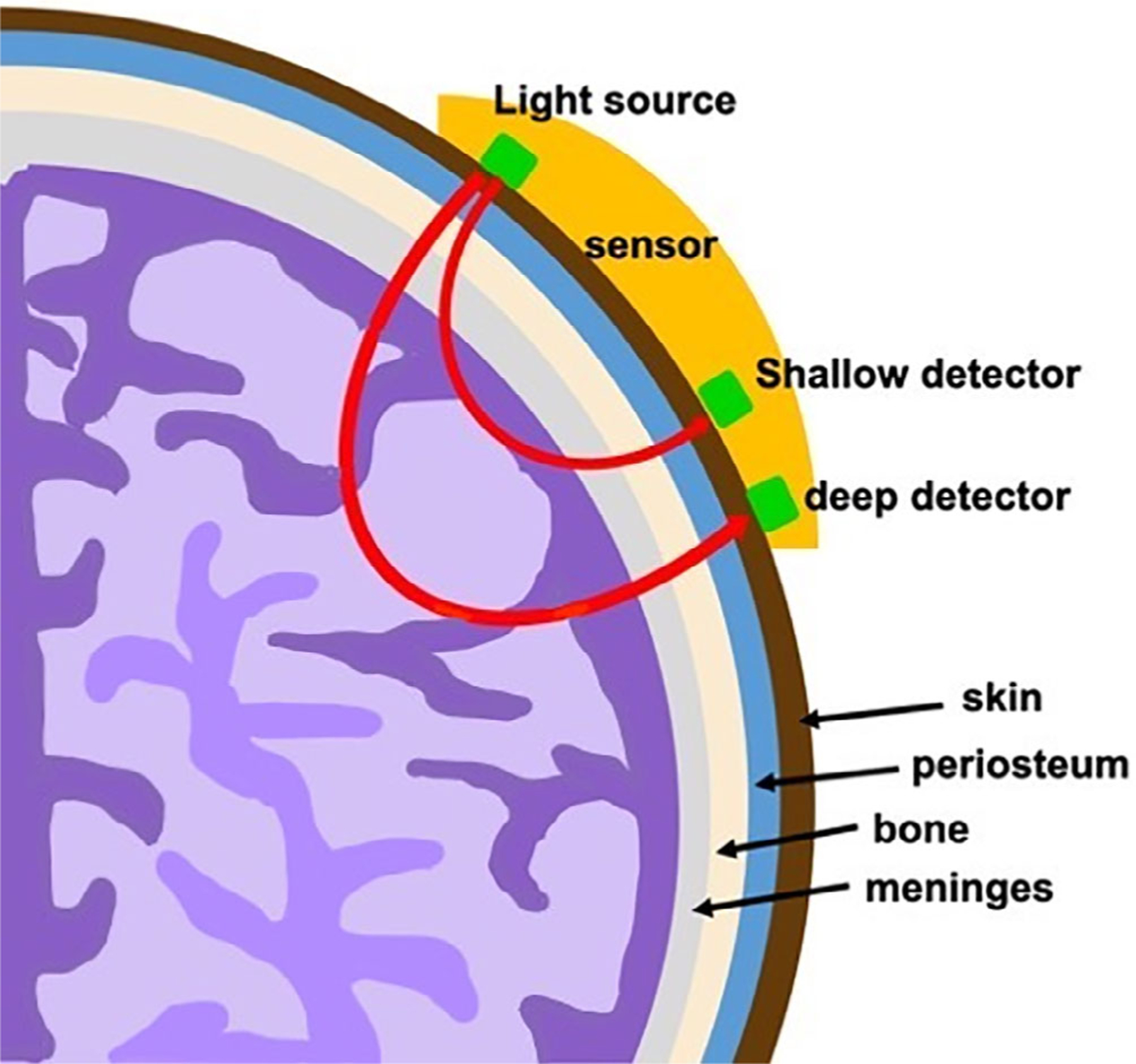
FIGURE 1.

Cerebral oximeter. Cerebral oxygenation is measured at the gray-white matter junction of the frontal lobes. Optodes are placed on each side of the patient’s head with deep and shallow signal detectors that are at different pre-determined distances from a light source; the signal from the shallow detector is subtracted from that of the deep detector to obtain the cerebral oxygenation measurement of the hemoglobin content only
