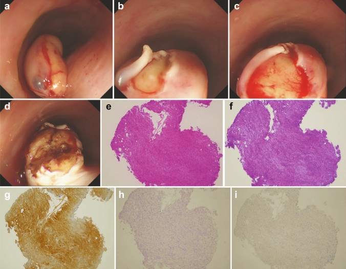
Fig. 3.
Mucosal incision-assisted biopsy procedure. a Submucosal injection with normal saline. (b, c) Longitudinal mucosal incision using DualKnife. d Biopsy of the tumor under direct vision. e H-E stain of the biopsy specimens showed small tumor cells with eosinophilic cytoplasm. f PAS stain revealed cytoplasm of tumor cells were rich with PAS-positive granules. g Immunohistochemical staining for S-100 protein showed tumor cells were positive for S-100 protein. h Immunohistochemical staining for KIT protein showed tumor cells were negative for KIT. i Immunohistochemical staining for Desmin showed tumor cells were negative for Desmin. H-E Hematoxylin–Eosin; PAS periodic acid Schiff

