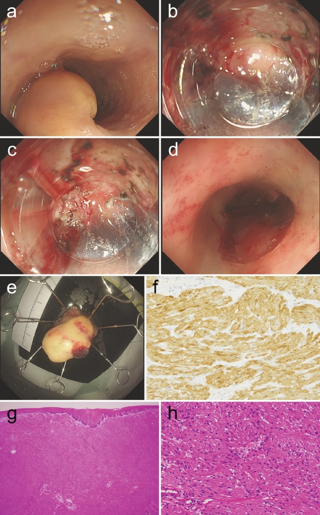
Fig. 4.

a A yellowish submucosal tumor at the middle intrathoracic esophagus. The tumor was completely dissected without any remnant lesion. (b, c) The tumor was resected by ESD under direct vision from the deeper side using a scissor-type device. d The wound from ESD showed no damage to the muscular layer. e Resected yellowish tumor. f Immunohistochemical staining for S-100 protein showed the tumor cells were positive for S-100. g H-E stain (× 40) of resected tumor showed tumor cells with eosinophilic cytoplasm proliferating in submucosa in solid alveolar form. h Under high magnification (× 400), tumor cells appeared as small cells with small uniform nuclei and eosinophilic and granule-rich cytoplasm. ESD Endoscopic submucosal dissection; H-E Hematoxylin–Eosin
