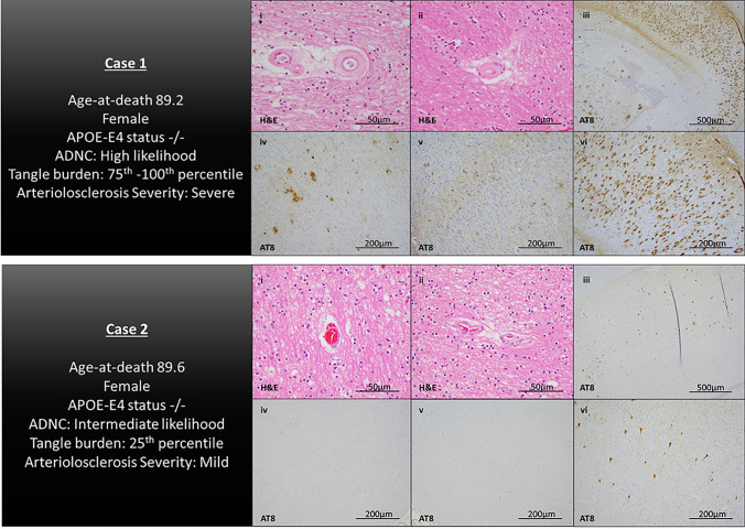
Fig. 5.
Illustrative cases. Histological sections from representative participants. Case 1 is a female participant, (age-at-death is 89.2 years), with a tau-tangle burden within the 75th–100th percentile and severe arteriolosclerosis pathology in watershed brain regions. Case 2 is a female participant (age-at-death is 89.6 years), with a tau-tangle burden within the 25th percentile and mild arteriosclerosis pathology in watershed brain regions. Images represent H&E-stained sections of the posterior watershed brain regions (i and ii) and AT8-stained sections for PHF-tau-tangle pathology in the CA1 subregion of the mid-hippocampus (iii and vi), midfrontal gyrus (iv), and inferior parietal cortex (v)

