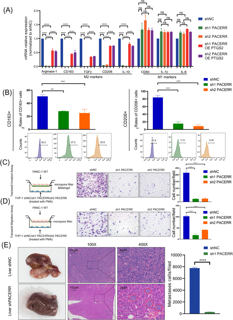
FIGURE 5.

PTGS2 antisense NF‐κB1 complex‐mediated expression regulator RNA (PACERR) knockdown hinders the M2 polarization and pro‐tumour functions of THP‐1‐derived tumour‐associated macrophages (TAMs). (A) Quantitative polymerase chain reaction (qPCR) analysis of the relative expression of M2 markers (Arginase‐1, CD163, TGFβ, CD206, and IL‐10) and an M1 marker (CD80, IL‐1β, IL‐6) in THP‐1‐derived TAMs after PACERR knockdown or prostaglandin‐endoperoxide synthase 2 (PTGS2) overexpression. THP‐1 cells were treated with PMA and cocultured with PANC‐1 cells for 2 days. Data are shown as the results from three independent experiments. (B) Flow cytometric analysis of the expression of M2 markers (CD163 and CD206) in THP‐1‐derived TAMs after PACERR knockdown. THP‐1 cells were treated with phorbol 12‐myristate 13‐acetate (PMA) and cocultured with PANC‐1 cells for two days. Data are shown as the results from three independent experiments. (C) Invasion capacity of PANC‐1 cells co‐cultured with THP‐1‐derived TAMs (shNC/ shPACERR). shNC means that cells were transfected in negative control plasmids. (D) Migration capacity of PANC‐1 cells co‐cultured with THP‐1‐derived TAMs (shNC/ shPACERR). (E) Representative images of liver metastasis and the number of metastatic cells in the PDAC mouse model, in which PANC‐1 cells mixed with TAMs (shNC/sh1 PACERR THP‐1) were injected into the spleens of BALB/c nude mice. Data are shown as the results from three independent experiments. * p < .05; ** p < .01; *** p < .001; **** p < .0001
