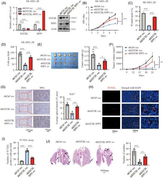
FIGURE 4.

EEF2K facilitates melanoma progression by targeting SPP1. (A) Real‐time PCR and Western blotting analysis of EEF2K and SPP1 in the indicated SK‐MEL‐28 cells. (B–D) Cell proliferative capacity measured by cell counting kit‐8 assay (B), cell migratory capacity quantified by wound healing assay (C) and cell invasive capacity identified by transwell assay (D) of the indicated SK‐MEL‐28 cells. (E and F) Tumour weight (E) and tumour volume (F) in the indicated groups. (G) Ki67 staining of the sectioned tumours to identify tumour cell proliferation in the indicated groups. (H and I) TUNEL assay to quantify apoptotic cells in xenografted tumours. (J) Metastatic nodules in lung indicated by haematoxylin–eosin staining. p‐Values were determined using one‐way ANOVA analysis in (A), (C–E), (G), (I) and (J). Two‐way ANOVA was performed in (B) and (F). *p < .05; **p < .01; ***p < .001
