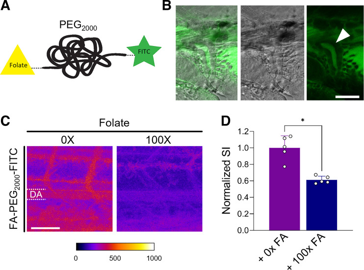
Figure 3.
Reabsorption of folate (FA) in the distal tubule. A: FA receptor 1 (folr1)-mediated distal tubular reabsorption was studied using a FA conjugate covalently modified with polyethylene glycol (PEG; molecular weight: 2,000 Da) and the fluorescent dye FITC. B: accumulation of in the lumen of the distal tubule 5 min postinjection of a 72-hpf zebrafish larva (ZFL). Scale bar = 30 µm. C: confocal microscopy image of the tail region of a 72-hpf ZFL 1 h after intravenous injection of a fluorescent-labeled FA-PEG2000-FITC derivative in the presence and absence of a 100-fold excess of native FA (100× FA). D: quantitative evaluation of the dorsal artery (DA) in C. Signal intensities (SI) were normalized to the mean of the control (no inhibitor, 0× FA). Values are means ± SD; n = 5. *P < 0.0001. Scale bar = 50 µm.

