
FIGURE 7.
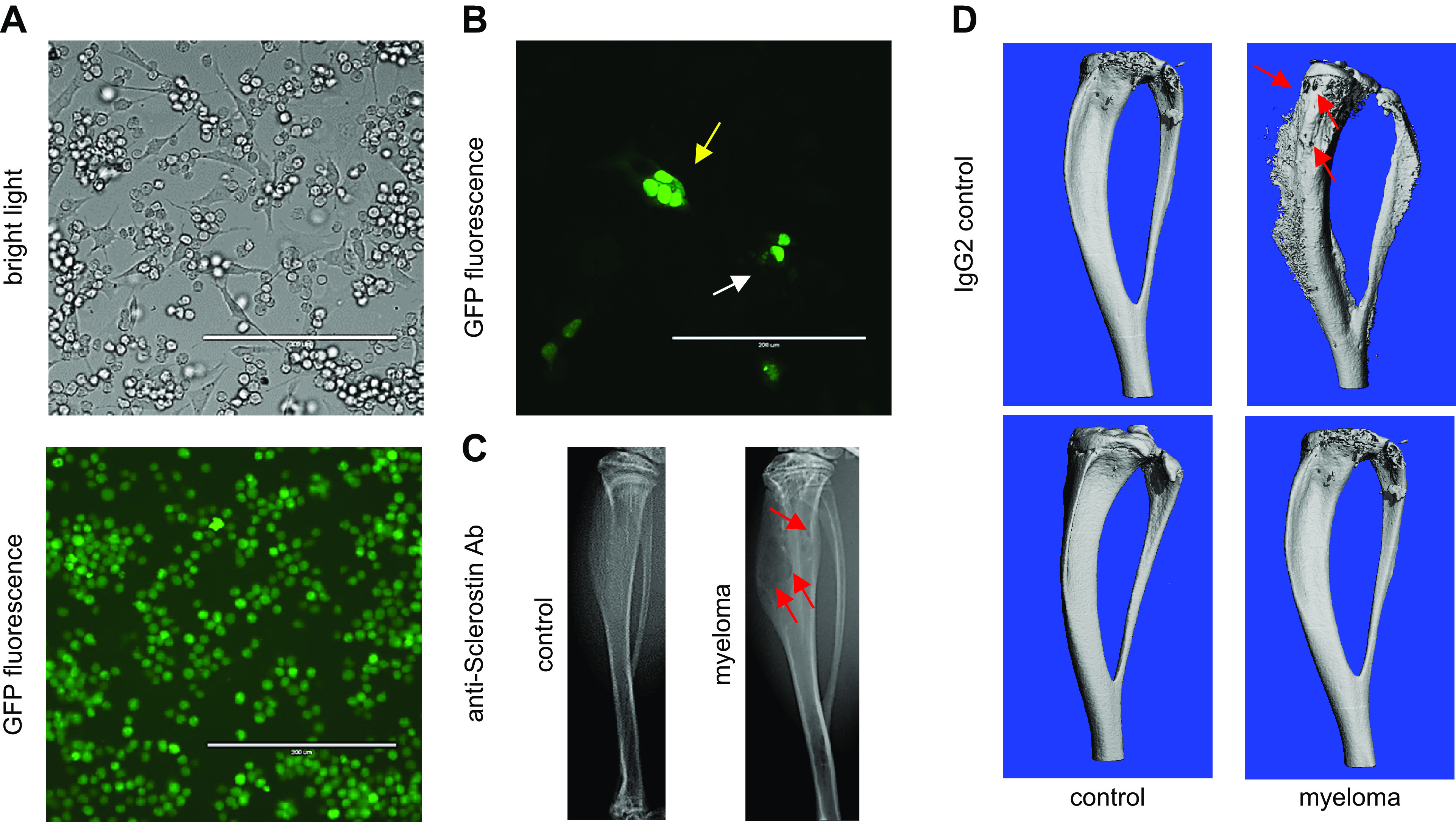
Osteocytes generate a microenvironment supportive for the growth of cancer cells and exacerbated bone resorption. A: cocultures of osteocytes and myeloma-green fluorescent protein (GFP) cancer cells enable the study of communication by cell-to-cell direct cell contact and by the exchange of soluble factors. Under these conditions, myeloma cells establish physical contact with the body and dendritic processes of osteocytes. B: osteocytes transfected with nuclear GFP cocultured with myeloma cancer cells. Fragmented (yellow arrow) and normal (white arrow) nuclei in MLO-Y4 osteocytes cocultured with MM1.S myeloma cells are displayed. C: murine intratibial injection of myeloma cells engrafts and produces multiple myeloma (MM) tumors in the bone marrow tumors that induce osteolytic lesions (red arrows) similar to those seen in myeloma patients. D: pharmacological blockade of the osteocyte-derived factor Sclerostin with a neutralizing antibody (anti-Sclerostin Ab) prevents the progression of the myeloma-induced bone disease in mice bearing myeloma tumors. Three-dimensional micro-computed tomography (μCT) reconstructions of C57BL/KaLwRijHsd bones bearing 5TGM1 myeloma cells are shown.