Figure 2(a-d).

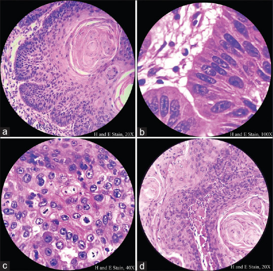
Histopathological image shows central squamous differentiation, peripheral palisading, increased mitotic activity and skeletal muscle infiltration of tumor cells

Histopathological image shows central squamous differentiation, peripheral palisading, increased mitotic activity and skeletal muscle infiltration of tumor cells