
Figure 6. Western blot analysis of MUC5AC and MUC5B collection by short washings in DMSO- and ETI-treated cells, followed by washings with a reducing agent.
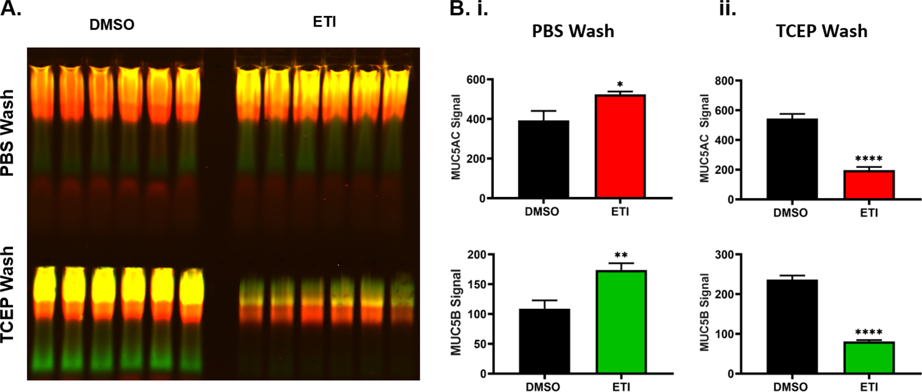
Following treatment for 3 days with 0.06% DMSO or 1μM VX-770, 2μM VX-445, and 3μM VX-661 (ETI), the apical surface of F508del HBE cells were washed with PBS for 15 min followed by an additional 1mM TCEP wash for 15 min. A.) Mucin western blot showing harvested MUC5AC (red) and MUC5B (green) by PBS (top) and additional 1mM TCEP (bottom) washings in DMSO- or ETI-treated cells. To stop the reaction, reduced samples were quenched with 1 mM iodoacetamide after 15min, n=6. B.) Graphs showing the intensity analysis of MUC5AC (top) and MUC5B (bottom) signals for PBS wash (i.) and additional TCEP wash (ii.). n=10. * P<0.05, ** P<0.01, **** P<0.0001, Student’s t-test. Error bars indicate SEM.