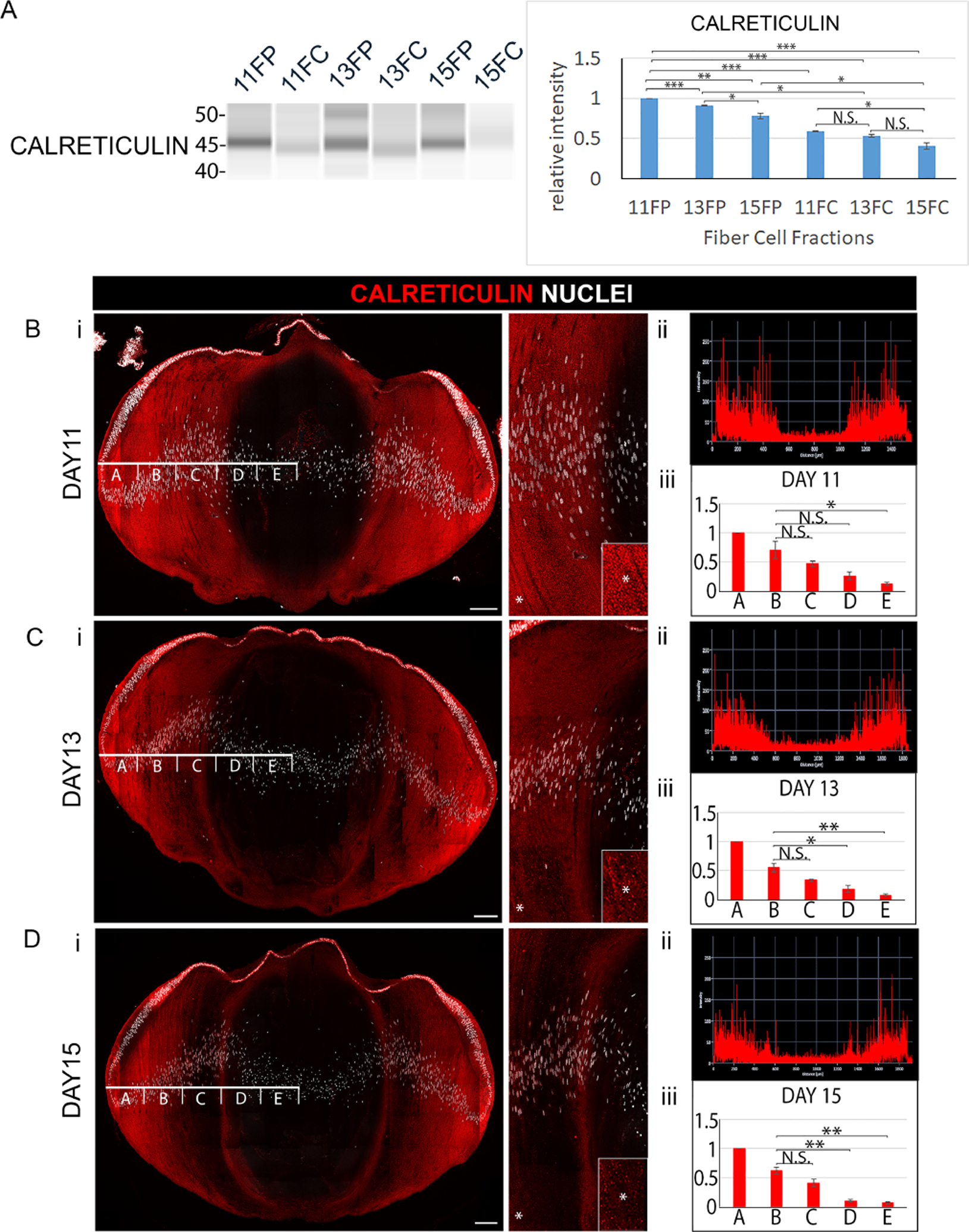
Figure 3. Elimination of endoplasmic reticulum (ER) to form the OFZ during development of the embryonic chick lens.

(A) Cortical fiber (FP) and central fiber (FC) regions of the chick embryo lens obtained by microdissection at E11, E13, and E15 were immunoblotted for the ER protein calreticulin. The quantification of immunoblots for calreticulin from a minimum of 3 independent studies is shown in the panel to the right. Lens cryosections were immunolabeled for calreticulin and co-labeled with DAPI at (Bi) E11, (Ci) E13, and (Di) E15. Each confocal image is shown on the left for the whole lens section and on the right as a zoomed in view in the region of the border of the cortical and central lens fiber cells. Inserts are of the regions denoted by an asterisk, shown at higher magnification. (Bii, Cii, Dii) Line scan analyses across the entire width of the lenses immunolabeled for calreticulin in Bi, Ci, Di, respectively. (Biii, Ciii, Diii) Bar graphs quantifying the fluorescence intensity from 3 independent studies over the left half of lenses as in Bi, Ci, Di, respectively, showing the staining intensities in the regions identified as A,B,C,D,E. These studies show the progressive removal of ER from fiber cells with lens development. Scale bars, 100 μm. Error bars represent S.E., *P ≤ 0.05, **P ≤ 0.01, and ***P ≤ .001, t test; N.S., not significant.
