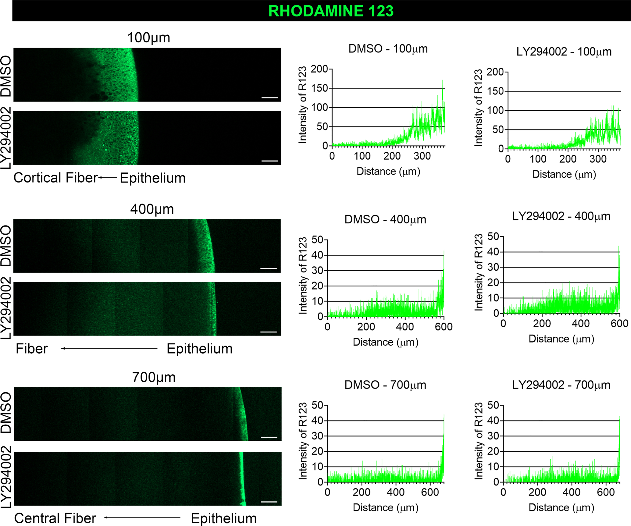
Figure 8. Inhibition of PI3K signaling does not impact mitochondrial membrane potential.

E12 lenses were treated for 24 hrs in organ culture with the pan-PI3K inhibitor, LY294002, or its vehicle DMSO and live labeled with Rhodamine 123 which is sequestered by active mitochondria and fluoresces green. The lens equatorial axis is positioned vertically with the anterior epithelium facing to the right and the lenses imaged by confocal microscopy at depths of 100 μm, 400 μm, and 700 μm. Fluorescence intensities were determined by line scan analyses and presented in graphical form to the right of the confocal images. The inhibition of PI3K signaling had no impact on mitochondrial membrane potential in the developing lens. Results are representative of 3 independent studies. Scale bar, 50 μm.
