FIGURE 3.

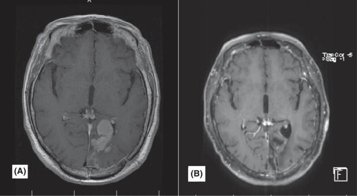
Case number 4. Post‐contrast axial brain MRI (A) brain MRI before treatment. Large mass in the left parietal lobe with avid non‐homogenous contrast enhancement. (B) after a follow‐up of 32 months. Gliomalacia in the left parietal lobe

Case number 4. Post‐contrast axial brain MRI (A) brain MRI before treatment. Large mass in the left parietal lobe with avid non‐homogenous contrast enhancement. (B) after a follow‐up of 32 months. Gliomalacia in the left parietal lobe