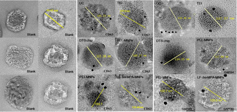
FIGURE 4.

Validation of exosome isolation from colon cancer CCM by immunogold electron microscopy. TEM micrographs of exosomes from six different isolation methods: UC, TEI, DTS chip, PLL‐MNPs, PEI‐MNPs, and LF‐bis‐MPA‐MNPs. Data for six different isolation methods were revealed by CD63‐labeling and GAPDH‐labeling. Blank (no labeling) corresponded to exosomes isolated by LF‐bis‐MPA‐MNPs. Exosome size ranged from 30 to 200 nm
