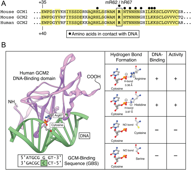
Figure 4.
Detailed analysis of the structural effect of R67 variants. A. Alignment of mouse and human GCM1 and GCM2. Conserved residues and conservatively substituted residues are drawn on a yellow background. Black dots (•) indicate DNA-contacting residues. Murine GCM1 arginine 62 and murine and human GCM2 arginine 67 are boxed. (B) Three-dimensional model of the GCM2 DNA-binding domain in complex with DNA. Ribbon representation of the GCM motif (pink) bound to its cognate DNA (green). The residue arginine 67 is shown in ball and stick presentation as is the DNA cytosine that it makes contact with. Expanded view of the structure of R67 and the substitutions: R67, H67, C67 and S67 shown in ball and stick format. In R67 and H67, the normal positive sidechain (oxygen atoms in blue) and negative DNA cytosine (hydrogen atoms in red) can form a hydrogen bond. In the presence of C67 or S67, the side chains cannot contribute to the formation of a hydrogen bond because they do not provide a positive charge. A full color version of this figure is available at https://doi.org/10.1530/EJE-21-0433.

 This work is licensed under a