Figure 5.
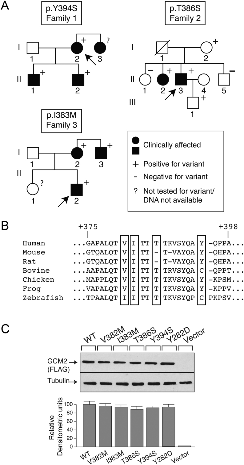
Detection of GCM2 variants in three kindreds with FIHP. (A) Pedigrees: clinical status is indicated by open symbols (unaffected) and solid symbols (affected). Proband is indicated by the arrow. The presence (+) or absence (−) of a GCM2 variant allele in tested family members is shown. Heterozygous c.1181A>C; p.Y394S (recurrent, Family 1); c.1156A>T; p.T386S (novel, Family 2); c.1149C>G; p.I383M (novel, Family 3). (B) The GCM2 protein sequences from diverse species were aligned as described in Materials and Methods. (C) Expression of WT and variant GCM2 proteins. Western blot analysis (top panel) of extracts of HEK293 cells that had been transfected with FLAG-tagged wild-type, or GCM2 variant constructs. β-tubulin was the loading control. Densitometric analysis of Western blot (lower panel).

 This work is licensed under a