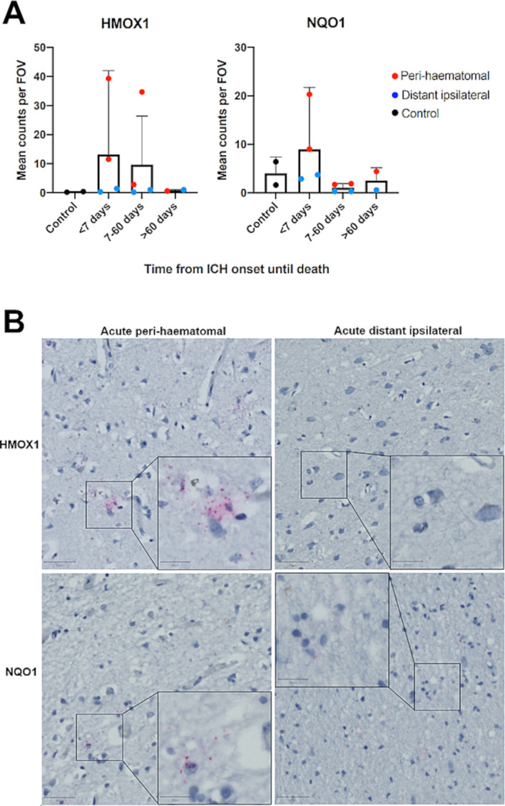
Figure 4.

(A) Mean (95% CI) in situ RNA hybridisation transcript counts per field of view (FOV) in sudden death controls versus ICH cases, by time of death after ICH symptom onset. Red points indicate perihaematomal and blue distant ipsilateral. Error bars: 95% CI. (B) Representative images of tissue from acute (<7 days from ICH onset until death) perihaematomal and distant ipsilateral tissue stained using fast red following RNA in situ hybridisation for HMOX1 or NQO1. Pink dots indicate transcripts haematoxylin counterstain. Scale bars=50 µm (main) and 20 µm (inset). HMOX1, haemoxygenase-1; ICH, intracerebral haemorrhage; NQO1, NAD(P)H dehydrogenase quinone 1.
