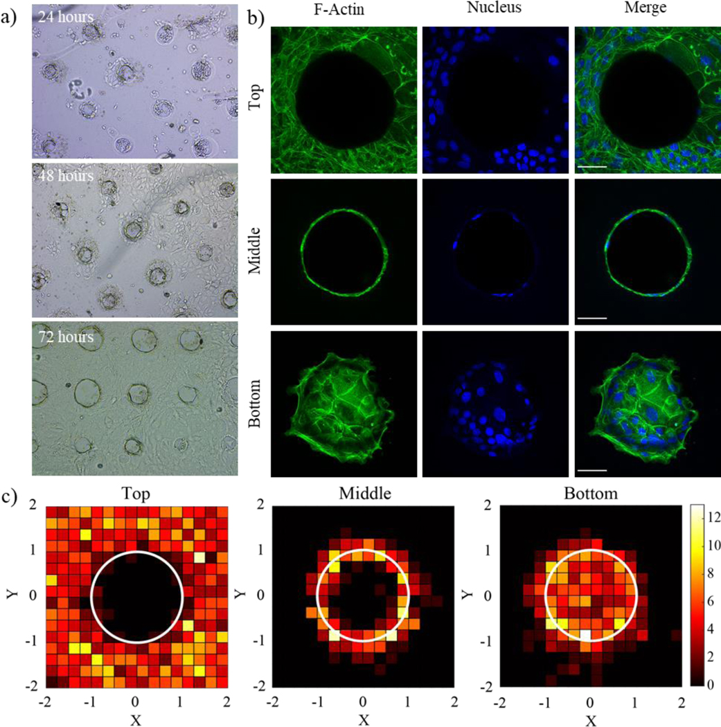
Figure 6.

a) Single intestinal stem cells were seeded onto patterned Matrigel gels and formed confluent monolayers over the course of three days. b) Immunostaining for F-actin (green) and nuclei (blue) shows the formation of a cell monolayer covering the patterned features. Scale bar is 50um. c) Frequency map images representing nuclear placement of ISCs along the top, middle, and bottom, of each well show even dispersion along the surfaces of the patterned well structures. X and Y image dimensions were normalized to the radius of each microwell which is represented by a white circle. The color bar represents the number of nuclei present in each region. N = 20 wells.
