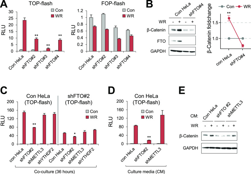
Figure 2. Loss of FTO suppresses canonical Wnt/β-Catenin signaling.
(A) HeLa cells stably expressing shRNAs targeting different sequences in FTO (#2, #3, #4; see the Materials and Methods section) were transfected with WNT signaling reporter (TOP-flash) or control reporter (FOP-flash) (n = 3). Relative Luciferase activities (RLU) were measured after 8 h of WNT stimulation. (B) Control HeLa cells or shFTO#4-expressing HeLa cells were stimulated with WR for 8 h and protein levels of β-Catenin and FTO were assessed by Western blot (n = 3). Note the stabilization of FTO protein by WNT signaling. (C) Control HeLa cells or shFTO#2-HeLa cells were transiently transfected with WNT reporter (TOP-flash) and co-cultured with cells expressing different shRNAs for 36 h as indicated (n = 3). Relative luciferase activities (RLU) were measured after 16 h of WNT stimulation. (D) Culture medium of HeLa cells transiently expressing WNT reporter (TOP-flash) was replaced with the medium harvested from control HeLa, shFTO#2-HeLa, or shMETTL3-HeLa before WNT stimulation for 16 h. RLU, Relative luciferase units (n = 3). (E) Cell lysates from Fig 2D were analyzed by Western blot for β-Catenin expression. GAPDH was used as a loading control for Western blot. WR, WNT3A, and R-Spondin1. Error bars are ± SEM. One-sided t test, **P < 0.01, *P < 0.05.

