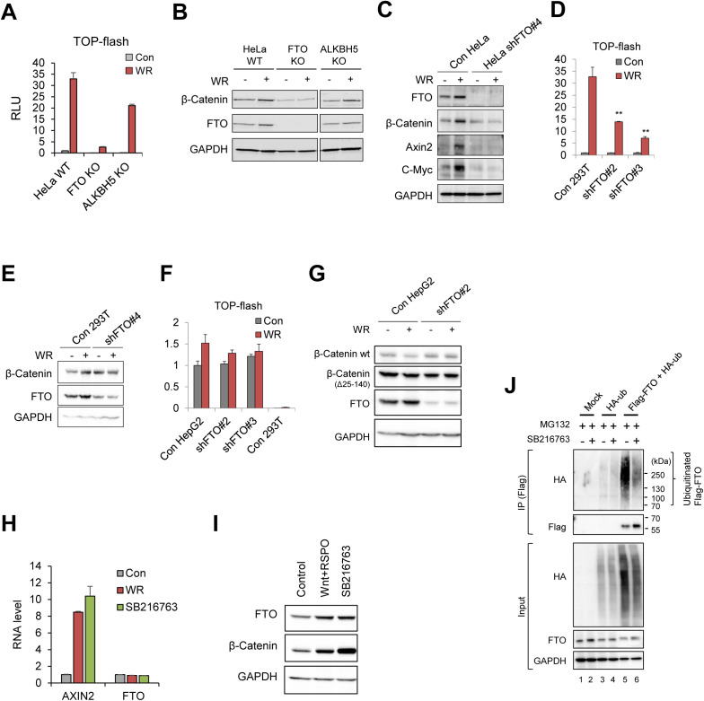
Figure S2. FTO is required for the canonical WNT/β-Catenin signaling.
(A) FTO KO and ALKBH5 KO HeLa cells were transiently transfected with WNT reporter (TOP-Flash) and stimulated with WR (WNT3a + RSPO1) for 12 h. Relative luciferase activities were measured (n = 6). (B) Protein levels of β-Catenin and FTO were measured by Western blot. GAPDH was used as a loading control. (C) Protein levels of FTO, β-Catenin, Axin2 and C-Myc were assessed in control or FTO-depleted HeLa cells treated with WNT3a and R-Spondin1 (WR) by Western blot. GAPDH was used as a loading control. (D) Control or FTO-depleted (shFTO#2 and shFTO#3, see the Materials and Methods section for details) 293T cells transiently expressing WNT reporter (TOP-flash) were stimulated with WNT (WR, WNT3a and R-Spondin1) for 16 h. Relative luciferase activities were measured (n = 3). (E) Control or FTO-depleted (shFTO#4) 293T cells were stimulated with WNT (WR, WNT3a, and R-Spondin1) for 16 h. Protein levels of β-Catenin and FTO were measured by Western blot. GAPDH was used as a loading control. (F) Control or FTO-depleted (shFTO#2 and shFTO#3) HepG2 cells transiently expressing WNT reporter (TOP-flash) were stimulated with WNT (WR, WNT3a, and R-Spondin) for 16 h. Relative luciferase activities were measured and compared with the results in control 293T cells (n = 3). (G) Control or FTO-depleted HepG2 cells were stimulated with WNT signaling and protein levels of β-Catenin and FTO were measured by Western blot. GAPDH was used as a loading control. Truncated form of β-Catenin (∼70 kD, Δ25-140) was detected in HepG2 cells as previously described (de La Coste et al, 1998). (H) FTO mRNA levels are unaffected by either WNT stimulation (WR) or GSK3 inhibition (SB216763). AXIN2 was used as positive control for WNT activity. GAPDH was used for the RT-qPCR normalization (n = 3). (I) Both WNT stimulation (WNT+RSPO) and GSK3 inhibition (SB216467) induce FTO protein stabilization along with β-Catenin stabilization. GAPDH was used as loading control. (J) Polyubiquitination of Flag-FTO in the presence of proteasomal inhibitor (MG132) and GSK3 inhibitor (SB216763). HeLa cells were transiently transfected with either mock, HA-ub alone or Flag-FTO together with HA-ub. After Flag immunoprecipitation, ubiquitinated FTO were assessed by Western blot using HA antibody. GAPDH was used as loading control. Error bars are ± SEM. One-sided t test. **P < 0.01.

