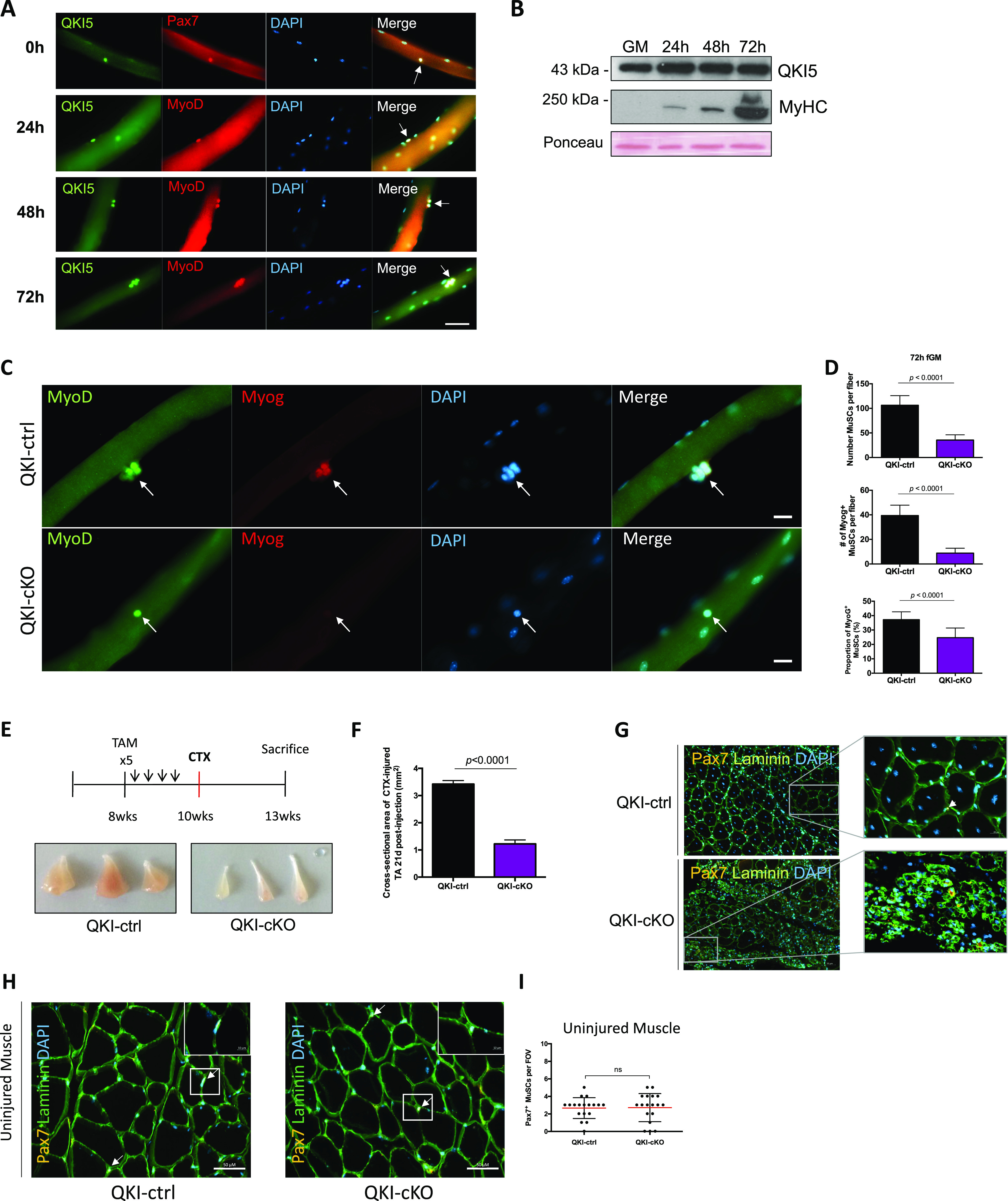
Figure 1. Mice with QKI-depleted muscle stem cells (MuSCs) exhibit reduced myogenic progenitors and defects in skeletal muscle regeneration.

(A) Representative immunofluorescence images of myofibers isolated from wild type mice and immunostained for QKI-5 (green), and co-stained with appropriate MuSC markers (Pax7 and MyoD; Red), counterstained with DAPI. Fibers were fixed immediately after isolation (0 h, quiescent MuSCs), and after 24, 48, and 72 h of culture. White arrows denote MuSCs. Scale white bar represents 50 μm. (B) Western blot of QKI-5 protein expression during a differentiation time course of primary mouse MuSCs (GM denotes growth media; 24–72 h represent time in differentiation media; MyHC is myosin heavy chain). Ponceau red was used to show equal protein loading. (C) Muscle fibers isolated from QKI-ctrl and QKI-cKO mice and cultured in fiber growth media for 72 h, stained with MyoD (green), Myogenin (red), and counterstained with DAPI (blue) and merged. Scale bar represents 10 μm. (C, D) Quantification of Myogenin-expressing MuSCs (upper panel) and total MuSCs (middle panel) from (C) (n = 3 biological replicates, minimum 1,000 cells quantified per condition, P < 0.0001, unpaired t test). Proportion of Myog + MuSCs from (C, lower panel) (n = 3 biological replicates, minimum 1,000 cells quantified per condition, P < 0.0001, unpaired t test). (E) Timeline of 4-hydroxytamoxifen injections (once daily for 5 d) to induce conditional QKI knockout in MuSCs of QKI2lox/2lox:Pax7CreERT2/+ or QKI2lox/2lox:Pax7+/+ as ctrl, followed by cardiotoxin injection in the tibialis anterior (TA) hindlimb muscle to induce muscle injury. 3 wk after injury, the mice were sacrificed and their TA muscles isolated (n = 6 biological replicates, three replicates depicted in bottom panels). (E, F) Quantification of cross-sectional area in square millimeter of TA muscles from QKI-ctrl and QKI-cKO mice in (E). (G) Representative immunofluorescence cross-sectional images of TA muscles 3 wk after cardiotoxin injury from QKI-ctrl and QKI-cKO. Laminin (green) stains muscle fiber edges, Pax7 (orange) indicates MuSCs, counterstained with DAPI (blue) (n = 6). (H) Cross-section of uninjured contralateral TA muscle of QKI-ctrl and QKI-cKO mice, laminin (green), Pax7 (orange), and DAPI (blue). Pax7+ MuSCs magnified in insets. Scale bars represent 50 μm. (H, I) Quantification of Pax7+ MuSCs per field of view in QKI-ctrl and QKI-cKO TA cross sections represented in (H).
