Figure 2.
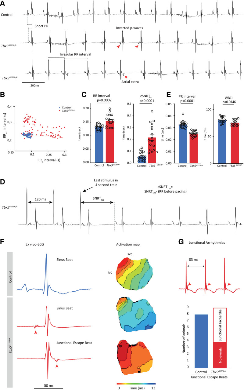
Transesophageal burst pacing (TEBP) of Tbx5G125R/+ mice shows atrial premature activation, shortened PR interval and ventricular premature activation. A, Typical example ECG traces of controls (n=19) and Tbx5G125R/+ mice (n=18). B, Scatterplot illustrating the beat-to-beat variability in RR interval of 1 control (blue) and 1 Tbx5G125R/+ mouse (red). C, Significant changes were observed in RR interval, RRsd (heart rate variability RRsd=SD of RRn–RRn+1), and SNRT of Tbx5G125R/+ mice (red). Genotypes were compared using Mann–Whitney U test; P values are given in the graphs. D, Typical example trace showing the analysis of cSNRT120. E, PR interval and WBCL are significantly shortened in Tbx5G125R/+ mice. Genotypes were compared using Mann–Whitney U test. F, Ex vivo typical example ECGs and corresponding activation maps (optical mapping) show atrial ectopic beats occurring during or after the QRS complex in response to a premature ventricular beat that originates in the atrioventricular junction in Tbx5G125R/+ mice. Red arrows indicate inverted premature beat (P-top). G, Junctional escape beats, not observed in 8 control mice, turn into junctional tachycardia in 55% (5/9) Tbx5G125R/+ mice. cSNRT120 indicates cyclic sinus node recovery time after 120-ms interval stimulation; ivc, inferior vena cava; SNRT, sinus node recovery time; svc, superior vena cava; TBX5, T-box transcription factor 5; and WBCL, Wenckebach cycle length.

