Fig. 5.
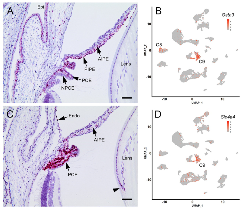
Expression of the cluster 9-enriched markers Gsta3 and Slc4a4 in anterior segment tissues of BALB/cJ mice. A. In situ hybridization with a Gsta3 probe reveals strong labeling (red) in the pigmented ciliary epithelial (PCE) layer but not in the non-pigmented ciliary epithelium (NPCE). The anterior and posterior iris pigment epithelium (AIPE and PIPE) and the corneal (Epi) and conjunctival epithelia are also labeled. B. Cluster analysis indicates that Gsta3 expression is largely restricted to cells in cluster 9 (C9) although some expressing cells are located in C8. C. Slc4a4 is strongly expressed in the PCE, although transcripts are also detected at a low level in the anterior iris pigment epithelium (AIPE) corneal endothelium (endo) and equatorial lens cells (arrowhead). D. Scatter plot showing that Slc4a4-expressing cells are largely restricted to C9. Scale bar A,C = 50 μm.

