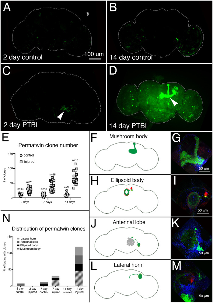
Figure 5.
Perma-twin lineage tracing demonstrates brain regeneration and appropriate targeting of axons following PTBI. To analyze neurogenesis after PTBI, we utilized the perma-twin lineage-tracing system (Fernandez-Hernandez et al. 2013). This system permanently labels dividing cells and their progeny with either GFP or RFP. Flies were reared at 17°C to keep the system off during development. Upon eclosion, F1 males carrying perma-twin transgenes were collected, injured and placed at 30°C to recover for either 2 or 14 days. (A) In 2-days uninjured controls, there are some GFP+ cells scattered throughout the brain. (B) At 14 days, there are relatively few GFP+ cells present in the control central brain. (C) In comparison, 2-day injured brains have more GFP+ cells that tend to cluster near the injury, (arrowhead). (D) At 14 days post-injury, there are large clones near the site of injury. Some of these clones have axons that project along the mushroom body tracts (arrowhead). Only the GFP channel is shown here; there were similar RFP+ clones in the PTBI samples. (E) The number of clones increases over time post-PTBI. Control uninjured brains (n = 13) have an average of 10 clones at 2 days while 2-day PTBI brains (n = 20) have an average of 23 clones (P < 0.00002). At 7 days, control brains had an average of 9 clones per brain (n = 18), while 7-day PTBI brains had an average of 39 clones per brain (n = 16) (P-value < 0.00000002). This is significantly more than the number of clones seen at 2 days post-injury (P-value < 0.0009). In 14-days control brains, there are an average of 10 clones per brain, which is not significantly different from the 2-day and 7-day controls. However, at 14 days post-PTBI, there are an average of 66 GFP+ clones, which is significantly more than either age-matched controls (P < 0.0000003) or 2-day post-PTBI brains (P-value < 0.0001). Error bars reflect SD. (F–M) PTBI stimulates clone formation in multiple regions in the brain. Panels on the left side are schematics of brain regions where large clones were found 14 days post-PTBI (A,H,J,L). Panels on the right show high magnifications of representative brains (G,I,K,M). Many 14-day brains had clones that projected to particular target areas. These included the mushroom body (MB) (F,G), the EB (H,I), the antennal lobe (AL) (J,K), and the lateral horn (LH) (L,M). (N) Both clone number and clone size increase with time post-PTBI. The proportions of brain regions with large clones were calculated at 2, 7, and 14 days in controls and injured brains. At 2 days, approximately 8% of control brains (n = 13) showed AL clones, while in 2-days injured brains (n = 20), there were no AL clones. In 7-days control brains (n = 18), 6% had AL and 6% had EB clones. At 7 days post-PTBI (n = 16), 6% of brains also had AL clones, 6% had EB clones, and 19% had large MB clones. At 14 days, control brains (n = 9) did not exhibit any specific areas with clones, while 47% of PTBI brains (n = 15) had MB clones, 20% of PTBI brains had AL clones, and 27% of PTBI brains had EB clones, and 27% had LH clones.

