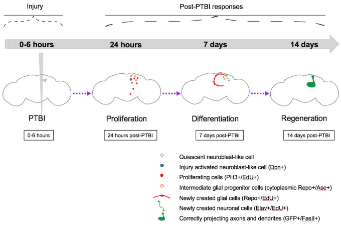
Figure 7.
Summary model for regeneration following PTBI. We propose that in young adult Drosophila there are quiescent NB-like cells within the central brain that lack expression of canonical NB genes. By 24 hours post-PTBI, the quiescent NB-like cells are activated, express NB genes, and have begun to proliferate. At both 4 and 24 hours post-PTBI, there is a wave of cell death as assayed using TUNEL. At 7 days, the proliferation rate is still high, and many of the new cells have adopted mature cell identities, becoming neurons or glia. At 10 days post-PTBI, there is no longer a difference in TUNEL+ cells between uninjured brains and injured brains, indicating that the wave of cell death has ended. Because the peaks of both cell death and proliferation occur at the same time post-injury, this could explain why there is not a significant increase in the number of EdU+ cells seen at 7 days compared to the number of PH3+ cells seen at 24 hours. However, by 10 days, cell death is back to control levels while proliferation has decreased but is still slightly above baseline levels. This could explain why there is an increase in the number of EdU+ between 7 and 14 days. At 14 days post-PTBI, there are large clones of new neurons with axons and dendrites correctly projecting to their respective target areas. Locomotor defects are also restored by 14 days, suggesting that adult Drosophila are able to regenerate functionally as well as structurally.

