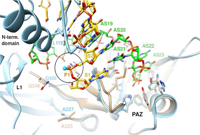
Figure 14.

Environment of the siRNA sense strand (S, yellow carbons) 5′ end paired opposite the siRNA antisense strand (AS, green carbons) and bound to the PAZ domain (residues 229 to 348 in human Ago2, light blue cartoon). The illustration shows an overlay of residues 227–346 from the structure of the human Ago1 PAZ domain (tan cartoon) bound to an siRNA-like duplex (PDB ID: 1SI3, (62)) and residues 229–348 from the structure of full-length human Ago2 in complex with miR-20a (PDB ID: 4F3T (61). The PAZ and N-terminal domains and linker L1 are labeled, selected S and AS siRNA and Ago residues are numbered, and the pro-Rp and pro-Sp oxygens of the first phosphate (circled) are highlighted as balls colored in pink and cyan, respectively.
