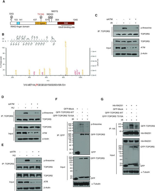
Figure 2.
TOPORS is phosphorylated at Thr515 in response to IR. (A) Schematic diagrams of the TOPORS protein domains with putative ATM phosphorylation sites indicated. (B) Determination of IR-induced phosphorylation sites in TOPORS by mass spectrometry. A peptide containing Thr515 phosphorylation is shown. HeLa cells transfected with control siRNA or ATM siRNA were treated with or without 5 Gy of IR (C) or 2 mM HU (D). Whole cell lysates were then subjected to immunoprecipitation using an anti-TOPORS antibody followed by immunoblotting using indicated antibodies. (E) HeLa cells transfected with control siRNA or ATR siRNA were treated with or without 2 mM HU. Whole cell lysates were then subjected to immunoprecipitation using an anti-TOPORS antibody followed by immunoblotting using indicated antibodies. (F) HEK293T cells transfected with control GFP vector, GFP-TOPORS-WT or GFP-TOPORS-T515A were treated with IR (5 Gy). Cell lysates were immunoprecipitated with anti-GFP antibody and subjected to immunoblot analysis with anti-phospho-threonine antibody. Asterisk indicates degradation products of GFP-TOPORS. (G) HA-RAD51-expressing HEK293T cells transfected with control GFP vector, GFP-TOPORS-WT or GFP-TOPORS-T515A were treated with 5 Gy of IR and subjected to immunoprecipitation and immunoblots as indicated. Asterisk indicates degradation products of GFP-TOPORS.

