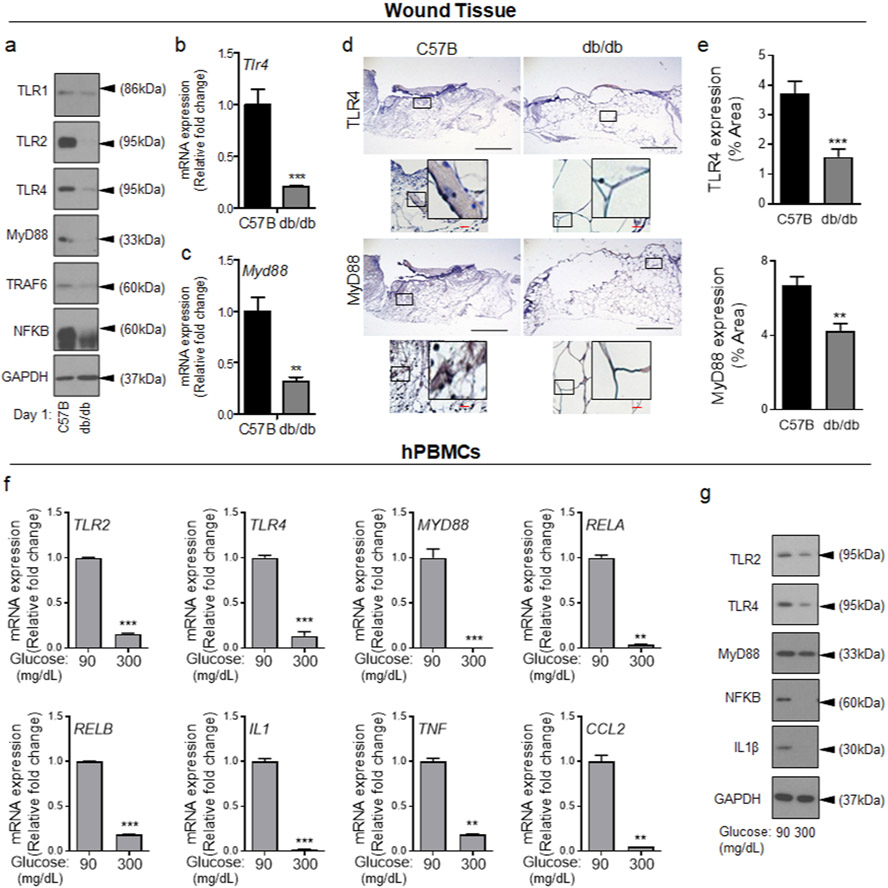
Figure 1. Toll-like receptor signaling is diminished during the acute phase wound healing in diabetic wound and in response to short-term exposure to HG.
C57B and db/db wounds were collected 24h after wounding and assessed for the expression of indicated genes by Western blotting (a), by mRNA analysis using RT-PCR (b-c), and by immunohistochemistry (d-e). Representative images are shown in (d) and the corresponding data in (e). (N=5 mice/group, ≥9 random fields/wound/mouse. Black scale bars=500μm; Red scale bars=50μm). (e-f) hPBMCs were exposed to 90mg/dl or 300 mg/dl for 1h and assessed for the expression of indicated genes by mRNA (f), or by Western blotting (g). Data are plotted as mean ± SEM. (Each experiment was repeated at least 2 times for (e) and at least 3 times for (f). *p<0.05, **p<0.01, ***p<0.001).

