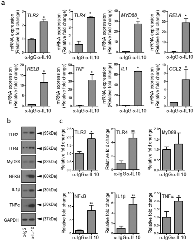
Figure 3. Blocking IL-10 signaling enhances TLR signaling and proinflammatory cytokines’ production in PBMCs in the presence of high glucose.

(a-c) hPBMCs were exposed to high glucose (300 mg/dl) for 1h in the presence of either α-IL-10 or α-IgG antibodies at 5μg/ml and assessed for mRNA levels of indicated genes using RT-PCR (a), or by protein analyses using Western blotting (b) and corresponding densitometer tabulated data for Western blots are in (c). All data are plotted as the mean ± SEM. (Each experiment was repeated at least 2 times for (a) and at least 3 times for (b-c); *p<0.05, **p<0.01, ***p<0.001. Pair-wise statistical analyses between groups were performed by unpaired Student’s t-test).
